Central Nervous System
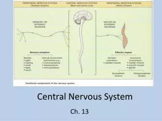
Central Nervous System . The nervous system has three specific functions : 1. Sensory input . Sensory receptors present in skin and organs respond to external and internal stimuli by generating nerve impulses that travel to the brain and spinal cord.

Central Nervous System
E N D
Presentation Transcript
The nervous system has three specific functions: 1. Sensory input. Sensory receptors present in skin and organs respond to external and internal stimuli by generating nerve impulses that travel to the brain and spinal cord. 2. Integration. The brain and spinal cord sum up the data received from all over the body and send out nerve impulses. 3. Motor output. The nerve impulses from the brain and spinal cord go to the effectors, which are muscles and glands. Muscle contractions and gland secretions are responses to stimuli received by sensory receptors
Characteristics of N.T • Neuron is the structural and functional unit • Neuron has central nucleus • Dendrites and axons are accessory part on neuron • No ability to regenerate ( if damage : fibrous tissue : neuroglaia ) • In N.T there is supporting cells their functions are : engolvment , support and CSF secretion
N.T consists of : • Cells • Fibers Fiber : axon ( short too long ) Group of axons nerve fibers Group of nerve fibers nerve Around N. fiber there is fat sheath called MYLINE accelerate impulses Function of N.T : control
Nervous Tissue nervous tissue is made up of just two principal types of cells: (1) neurons, also called nerve cells, which transmit nerve impulses; (2) neuroglia, which supports and nourishes neurons
Neuron Structure Neurons vary in appearance, but all of them have just three parts: a cell body, dendrite(s), and an axon., the cell body contains the nucleus as well as other organelles.
In motor neurons,the dendrites are the many short extensions that receive signals from sensory receptors or other neurons. • At the dendrites, signals can result in nerve impulses that are then conducted by an axon. • The axon is the portion of a neuron that conducts nerve impulses.
Any long axon is also called a nerve fiber. • Long axons arecovered by a white myelin sheath formed from the membranes of tightly spiraled neuroglia. • In the PNS, a neuroglialcell called a neurolemmocyte (Schwann cell) performs this function, leaving gaps called neurofibril nodes (nodes of Ranvier). • Another type of neuroglial cell performs a similar function in the CNS.
Types of Neurons Neurons can be classified according to their function and shape. Motor neurons take nerve impulses from the CNS to muscles or glands. Motor neurons are said to be multipolar because they have many dendrites and a single axon Motor neurons cause muscle fibers to contract or glands to secrete, and therefore they are said to innervate these structures.
Sensory neurons take nerve impulses from sensory receptors to the CNS. The sensory receptor, which is the distal end of the long axon of a sensory neuron, may be as simple as a naked nerve ending (a pain receptor), or it may be a part of a highly complex organ, such as the eye or ear. Almost all sensory neurons have a structure that is termed unipolar
Bipolar neurons are located in some sensory organs, such as in the retina of the eye and in the nasal cavity.
Neuroglia of the CNS Neuroglia are far more numerous than neurons and account for more than half the brain’s weight. • They are the major supporting cells in the CNS, • participate in the formation of a permeability barrier between the blood and the neurons, • phagocytize foreign substances, • produce cerebrospinal fluid, • form myelin sheaths around axons.
There are four types of CNS neuroglial cells and each has unique structural and functional characteristics
Ependymal : cells line the ventricles (cavities) of the brain and the central canal of the spinal cord. • The free surface of the ependymal cells frequently has patches of cilia that assist in moving cerebrospinal fluid through the cavities of the brain
Microglia are specialized macrophages in the CNS that become mobile and phagocytic in response to inflammation, and they phagocytize necrotic tissue, microorganisms, and foreign substances that invade the CNS
Oligodendrocytes have cytoplasmic extensions that can surround axons. If the cytoplasmic extensions wrap many times around the axons, they form myelin sheaths. A single oligodendrocyte can form myelin sheaths around portions of several axons
The Brain We will discuss the parts of the brain with reference to the :cerebrum, the diencephalon, the cerebellum, and the brain stem. The brain’s four ventricles are called :the two lateral ventricles, the third ventricle, and the fourth ventricle. It will be helpful for you to associate the cerebrum with the two lateral ventricles, the diencephalon with the third ventricle, and the brain stem and the cerebellum with the fourth ventricle
The Cerebrum • The cerebrum is the largest portion of the brain in humans. • The cerebrum is the last center to receive sensory input and carry out integration before commanding voluntary motor responses. • It communicates with and coordinates the activities of the other parts of the brain. The cerebrum carries out the higher thought processes required for learning and memory and for language and speech.
The Cerebral Hemispheres: The cerebrum has two halves called the left and right cerebral hemispheres A deep groove, the longitudinal fissure, divides the left and right cerebral hemispheres. Still, the two cerebral hemispheres are connected by a bridge of white matter within the corpus callosum
Gyrus …gyri • Sulcus …sulci
Diencephalon Enclosed by the cerebral hemispheres Located above the brain stem Has three major structure: • Thalamus :enclosed third ventricle. • Hypothalamus: regulathion of the body temperature, emotions, thirst, appetite, pain, sex, pleasure and regulate the pitutary gland • epithalamus : forms the roof of the third ventricle
Brain stem Has three major structure: • Midbrain : vission,hearing • Pons : breathing control • Medulla oblongata: contains centers that control heart rate ,blood pressure,breathing swallowing, vomiting
Cerebellum Play a central role in the control of the timing of the skelelal muscle activity and controls our balance and equilibrium