Central Nervous System
Central Nervous System. Ch. 13. Introduction. CNS consists of brain & spinal cord The brainstem connects the brain to spinal cord Communication to PNS is by way of spinal cord. Arachnoid Mater: “spider-web like” Space contains cerebrospinal fluid (CSF) Pia Mater: “faithful mother”

Central Nervous System
E N D
Presentation Transcript
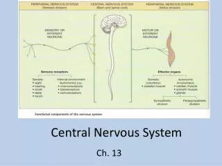
Central Nervous System Ch. 13
Introduction • CNS consists of brain & spinal cord • The brainstem connects the brain to spinal cord • Communication to PNS is by way of spinal cord
Arachnoid Mater: “spider-web like” Space contains cerebrospinal fluid (CSF) Pia Mater: “faithful mother” Encapsulates blood vessels Meninges • Membranes of CNS • Protect CNS • Made up of 3 layers: • Dura Mater: “tough mother” • Venous sinuses • Falx
Gray matter White matter
Ventricles & Cerebrospinal Fluid • 4 ventricles • Interconnected cavities w/in cerebral hemispheres & brain stem • Continuous with central canal of spinal cord • Filled with CSF
Ventricles: Lateral ventricles(2) Known as 1st & 2nd ventricles 3rd ventricle 4th ventricle Interventricular foramen Cerebral aqueduct Ventricles & CSF
Cerebrospinal Fluid • Secreted by the choroid plexus • Circulates in ventricles, central canal of spinal cord, & subarachnoid space • Completely surrounds the brain & spinal cord
Cerebrospinal Fluid • Excess or wasted CSF is absorbed by arachnoid villi • Clear fluid similar to blood plasma • Vol = about 120 mL • Nutritive & protective • Helps maintain stable ion concentrations in CNS
CSF Pressure remains relatively constant Infection, tumor or blood clot can incr pressure in ventricles by interfering with fluid’s circulation Can cause collapsed blood vessels, injured brain tissue CSF Pressure
CSF Pressure • Lumbar puncture (spinal tap) – bt 3rd & 4th lumbar vertebrae; measures pressure; removes fluid to look for blood cells
CSF Pressure • Temporary drain can relieve pressure • Fetus/infant – can lead to hydrocephalus • Shunt redirects fluid to digestive tract
Unhealthy CSF Healthy CSF – Lauren Shaw <3
Brain • Functions of Brain • Interprets sensations • Determines perceptions • Stores memory • Reasoning • Makes decisions • Coordinates muscular movements • Regulates visceral activities • Determines personality
Major Parts of the Brain • Cerebrum • Frontal lobes • Parietal lobes • Occipital lobes • Temporal lobes • Insula
Major Parts of the Brain • Diencephalon • Cerebellum • Brainstem • Midbrain • Pons • Medulla oblongata
Corpus callosum Connects cerebral hemispheres (a commissure) Gyri Bumps or convolutions Sulci Grooves in gray matter Central Sulcus of Rolando Structure of the Cerebrum
Structure of Cerebrum • Fissures • Longitudinal: separates the cerebral hemispheres • Transverse: separates cerebrum from cerebellum • Lateral fissure of Sylvius
4 lobes bilaterally: Frontal lobe Parietal lobe Temporal lobe Occipital lobe and Insula aka “Island of Reil” Lobes of Cerebrum
Functions of Cerebrum • Interpreting impulses • Initiating voluntary movements • Storing info as memory • Retrieving stored info • Reasoning • Seat of intelligence and personality
Functional Regions of Cerebral Cortex • Cerebral Cortex • Thin layer of gray matter that constitutes the outermost portion of cerebrum
Functional Regions of Cerebral Cortex • Contains 75% of all neurons in nervous system
Sensory Areas (post-central sulcus) • Cutaneous sensory area • Parietal lobe • Interprets sensations on skin
Visual area Occipital lobe Interprets vision Auditory area Temporal lobe Interprets hearing Sensory Areas (post-central sulcus)
Sensory area for smell Arises from centers deep within cerebrum Sensory area for speech Wernicke’s area Sensory Areas (post-central sulcus)
Gnostic area – stg for complex memory patterns assoc w/sensation; dmg causes imbecilic behavior due to inability to interpret any sensation Gustatory cortex – sense of taste Sensory Areas (post-central sulcus)
Affective language area – opp of Broca’s, nonverbal emotional components of language; expression of emotions associated with speech (“lilt” of one’s voice) Sensory Areas (post-central sulcus)
Association fibers – connect lobes • Projection fibers – connect cortex with lower brain or cord centers
Association Areas • Regions that are not primary motor or primary sensory areas • Widespread throughout cerebral cortex
Association Areas • Analyze/interpret sensory experiences • Provide memory, reasoning, verbalization, judgment, emotions
Association Areas • Frontal lobe assoc areas • Concentrating • Planning • Complex problem solving
Association Areas • Parietal lobe assoc areas • Understanding speech • Choosing words to express thought
Association Areas • Temporal lobe assoc areas • Interpret complex sensory experiences • Store memories of visual scenes, music, & complex patterns
Association Areas • Occipital lobe assoc areas • Analyze & combine visual images w/other sensory experiences
Motor Areas (pre-central sulcus) • Primary motor areas • Frontal lobes • Control voluntary muscles
Motor Areas (pre-central sulcus) • Broca’s Area • Anterior to primary motor cortex • Usually in left hemisphere • Controls muscles needed for speech
Motor Areas (pre-central sulcus) • Frontal eye field • Above Broca’s area • Controls voluntary movements of eyes & eyelids
Hemisphere Dominance • Left hemisphere is dominant in most individuals – 91% • Related to right-handedness
Hemisphere Dominance Dominant hemisphere controls: • Speech • Writing • Reading • Verbal skills • Analytical skills • Computational skills
Hemisphere Dominance • Non-dominant hemisphere controls: • Non-verbal tasks; • Holistic interpretation • Motor tasks • Spatial relations • Understanding/interpreting musical, artistic, visual patterns • Imagination & insight • Provides emotional & intuitive thought processes
Lateralization develops with age • females have more communication between hemispheres (corpus callosum thicker posteriorly)
Brain lesions • parietal lobe • contralateral neglect syndrome • temporal lobe • agnosia - inability to recognize objects • prosopagnosia - inability to recognize faces • frontal lobe • problems with personality (inability to plan and execute appropriate behavior)
Lobotomy of Phineas Gage • Ventromedial region of both frontal lobes • Personality change • irreverent, profane • Prefrontal cortex functions • planning, moral judgment, and emotional control
Alzheimer Disease • 100,000 deaths/year • 11% of population over 65; 47% by age 85 • Memory loss for recent events, moody, combative, lose ability to talk, walk, and eat