Download
1 / 38
380 likes | 460 Vues
Explore the functional anatomy of the respiratory system including the major organs like the nose, pharynx, larynx, and lungs. Learn about the process of respiration, gas exchange, and the role of respiratory muscles in ventilation.

E N D
Respiration • Pulmonary ventilation (breathing):movement of air into and outof the lungs • External respiration: O2 and CO2exchange between the lungsand the blood • Transport: O2 and CO2in the blood • Internal respiration: O2 and CO2exchange between systemic bloodvessels and tissues Respiratory system Circulatory system
Respiratory System: Functional Anatomy • Major organs • Nose, nasal cavity, and paranasal sinuses • Pharynx • Larynx • Trachea • Bronchi and their branches • Lungs and alveoli
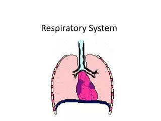
Nasal cavity Oral cavity Nostril Pharynx Larynx Left main (primary) bronchus Trachea Carina of trachea Right main (primary) bronchus Left lung Diaphragm Right lung Figure 22.1
Functional Anatomy • Respiratory zone: site of gas exchange • Microscopic structures: respiratory bronchioles, alveolar ducts, and alveoli • Conducting zone: passageway to gas exchange sites • Includes all other respiratory structures • Respiratory muscles: diaphragm and other muscles that promote ventilation
Nose • Functions • Provides an airway for respiration • Moistens and warms the entering air • Filters and cleans inspired air • Serves as a resonating chamber for speech • Houses olfactory receptors • Mucosa • Pseudostratified ciliated columnar epithelium • Cilia move contaminated mucus posteriorly to throat
Paranasal Sinuses • In frontal, sphenoid, ethmoid, and maxillary bones • Lighten the skull and help to warm and moisten the air
Pharynx • Muscular tube that connects to the • Nasal cavity and mouth superiorly • Larynx and esophagus inferiorly • From the base of the skull to the level of the sixth cervical vertebra Nasopharynx Oropharynx Laryngopharynx (b) Regions of the pharynx Figure 22.3b
Larynx • Attaches to the hyoid bone and opens into the laryngopharynx • Continuous with the trachea • Functions • Provides an airway • Routes air and food into proper channels • Voice production Hyaline cartilage keeps larynx and trachea open Epiglottis: elastic cartilage; covers the larynx during swallowing
Epiglottis Body of hyoid bone Thyrohyoid membrane Thyrohyoid membrane Fatty pad Cuneiform cartilage Vestibular fold (false vocal cord) Corniculate cartilage Arytenoid cartilage Thyroid cartilage Arytenoid muscles Vocal fold (true vocal cord) Cricoid cartilage Cricothyroid ligament Cricotracheal ligament Tracheal cartilages (b) Sagittal view; anterior surface to the right Figure 22.4b
Base of tongue Epiglottis Vestibular fold (false vocal cord) Vocal fold (true vocal cord) Glottis Inner lining of trachea Cuneiform cartilage Corniculate cartilage (a) Vocal folds in closed position; closed glottis (b) Vocal folds in open position; open glottis Figure 22.5
Voice Production • Speech: intermittent release of expired air while opening and closing the glottis • Pitch is determined by the length and tension of the vocal cords • Loudness depends upon the force of air • Chambers of pharynx, oral, nasal, and sinus cavities amplify and enhance sound quality • Sound is “shaped” into language by muscles of the pharynx, tongue, soft palate, and lips
Trachea • Windpipe Mucosa: ciliated pseudostratified epithelium with goblet cells Rings of Hyaline Cartilage
Posterior Mucosa Esophagus Submucosa Trachealis muscle Seromucous gland in submucosa Lumen of trachea Hyaline cartilage Adventitia Anterior (a) Cross section of the trachea and esophagus Figure 22.6a
Conducting Zone Structures • Trachea right and left main (primary) bronchi • Each main bronchus enters one lung • Right main bronchus is wider, shorter, & more vertical than the left Trachea Superior lobe of left lung Left main (primary) bronchus Superior lobe of right lung Lobar (secondary) bronchus Segmental (tertiary) bronchus Middle lobe of right lung Inferior lobe of left lung Inferior lobe of right lung Figure 22.7
Conducting Zone Structures • From bronchi through bronchioles, structural changes occur • Cartilage rings give way to plates; cartilage is absent from bronchioles • Epithelium changes from pseudostratified columnar to cuboidal; cilia and goblet cells become sparse • Relative amount of smooth muscle increases
Respiratory Zone • Respiratory bronchioles, alveolar ducts, alveolar sacs (clusters of alveoli) • ~300 million alveoli account for most of the lungs’ volume and are the main site for gas exchange Alveoli Alveolar duct Respiratory bronchioles Alveolar duct Terminal bronchiole Alveolar sac (a)
Three Types of Cells found in alveoli - Type I Cells – simple squamous cells; allow for gas diffusion - Surfactant Cell (Type II) – produce an oily secretion; it reduces surface tension so lungs don’t collapse - Alveolar Macrophages – Clear/swallow 2 million dust cells out of your lungs/hour. Type II Cell Macrophage
Red blood cell Nucleus of type I (squamous epithelial) cell Alveolar pores Capillary O2 Capillary Type I cell of alveolar wall CO2 Alveolus Macrophage Alveolus Endothelial cell nucleus Alveolar epithelium Fused basement membranes of the alveolar epithelium and the capillary endothelium Respiratory membrane Red blood cell in capillary Alveoli (gas-filled air spaces) Type II (surfactant- secreting) cell Capillary endothelium (c) Detailed anatomy of the respiratory membrane Figure 22.9c
Right lung Left lung Right superior lobe (3 segments) Left superior lobe (4 segments) Right middle lobe (2 segments) Left inferior lobe (5 segments) Right inferior lobe (5 segments) Figure 22.11
Blood Supply • Pulmonary circulation (low pressure, high volume) • Pulmonary arteries deliver systemic venous blood • Branch profusely, along with bronchi • Feed into the pulmonary capillary networks • Pulmonary veins carry oxygenated blood from respiratory zones to the heart
Blood Supply • Systemic circulation (high pressure, low volume) • Bronchial arteries provide oxygenated blood to lung tissue • Arise from aorta and enter the lungs • Supply all lung tissue except the alveoli • Pulmonary veins carry most venous blood back to the heart
Mechanics of Breathing • Pulmonary ventilation consists of two phases • Inspiration: gases flow into the lungs • Expiration: gases exit the lungs Caused by changes between atmospheric gas pressure (14.7 psi) and gas pressure in the lungs
Atmospheric pressure Movement of air always flows from higher gas pressure to lower gas pressure Thoracic wall 756 Intrapleural pressure 756 mm Hg (–4 mm Hg) 760 Intrapulmonary pressure 760 mm Hg (0 mm Hg) Lung Diaphragm Figure 22.12
Pulmonary Ventilation • Inspiration and expiration • Mechanical processes that depend on volume changes in the thoracic cavity • Volume changes pressure changes • Pressure changes gases flow to equalize pressure
Boyle’s Law • The relationship between the pressure and volume of a gas • Used to explain the mechanics of breathing • Under constant temperature, Pressure (P) varies inversely with volume (V): P1V1 = P2V2 P=1/V • Lungs can change volume, so air pressure in lungs will also change
Inspiration • An active process • Inspiratory muscles contract • Lungs are stretched and volume increases • Air pressure in lungs drops • Air flows into the lungs
Changes in lateral dimensions (superior view) Changes in anterior- posterior and superior- inferior dimensions Sequence of events 1 Inspiratory muscles contract (diaphragm descends; rib cage rises). Ribs are elevated and sternum flares as external intercostals contract. Thoracic cavity volume increases. 2 External intercostals contract. 3 Lungs are stretched; intrapulmonary volume increases. Intrapulmonary pressure drops. 4 5 Air (gases) flows into lungs down its pressure gradient until intrapulmonary pressure is 0 (equal to atmospheric pressure). Diaphragm moves inferiorly during contraction. Figure 22.13 (1 of 2)
Expiration • Quiet expiration is normally a passive process • Inspiratory muscles relax • Thoracic cavity volume decreases • Elastic lungs recoil and volume inside lungs decreases • Pressure inside lungs increases • Air flows out of the lungs • Note: forced expiration is an active process: it uses abdominal and internal intercostal muscles
Changes in lateral dimensions (superior view) Changes in anterior- posterior and superior- inferior dimensions Sequence of events 1 Inspiratory muscles relax (diaphragm rises; rib cage descends due to recoil of costal cartilages). Ribs and sternum are depressed as external intercostals relax. 2 Thoracic cavity volume decreases. 3 Elastic lungs recoil passively; intrapulmonary volume decreases. External intercostals relax. 4 Intrapulmonary pres- sure rises. Diaphragm moves superiorly as it relaxes. 5 Air (gases) flows out of Lungs. Figure 22.13 (2 of 2)
Lung Volumes Tidal volume Inspiratory reserve volume Expiratory reserve volume Vital Capacity Residual Volume Total Lung Capacity
Tidal volume – normal breath in and out Vital Capacity – total amount of air you can forcefully inspire and expire in one respiratory cycle
Physical Factors Influencing Pulmonary Ventilation • Inspiratory muscles consume energy to overcome three factors that hinder air passage and pulmonary ventilation • Airway resistance • Lung compliance • Lung Elasticity
Airway Resistance • As airway resistance rises, breathing movements become more strenuous • Severely constricting or obstruction of bronchioles • Can prevent life-sustaining ventilation • Can occur during acute asthma attacks and stop ventilation • Epinephrine dilates bronchioles and reduces air resistance
Air Resistance • Friction in the respiratory passageways, decreases the flow of gases. • I.e. Asthma attack, constriction of bronchiole tubes • Accumulation of mucus (infections) • Cystic Fibrosis - mutation on chromosome #7 where lungs produce too much mucus • Leads to poor gas exchange since mucus blocks diffusion of gases.
Lung Elasticity The ability of the lung to recoil I.e. Emphysema Air sacs enlarge and lose their elasticity; lungs remain over inflated Lung Compliance • The ease at which lungs stretch • Decrease in Lung compliance occurs when: • Smaller passageways are blocked • Diminished by • Nonelastic scar tissue (fibrosis) • Reduced production of surfactant • Decreased flexibility of the thoracic cage
Lung Elasticity • The lungs’ ability to recoil • I.e. Emphysema • Air sacs enlarge and lose their elasticity; lungs remain over inflated


