Cell Division
Explore the intricate processes of cell division through mitosis and meiosis, the cell cycle phases, and the roles of microtubules, centrioles, and chromosomes. Understand DNA synthesis, ploidy numbers, and the critical functions of microtubules during cell division. Follow the dynamic changes in centrioles throughout the cell cycle phases and learn about the distinct stages of mitosis - from prophase to telophase - and cytokinesis. Delve into the details of each phase and gain insights into chromosome movements, spindle formation, and the separation of daughter cells.

Cell Division
E N D
Presentation Transcript
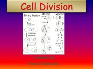
Cell Division Mitosis and Meiosis
Cell Cycle Phases • G0: “G-Naught” • Cells that are not actively preparing to divide • Terminal state for differentiated cells • Or Temporary resting stage. • S: “Synthesis” • New DNA is synthesized via DNA Replication G0 Interphase Interphase InterphaseInterphase Interphase Interphase • G1: “First Gap” • Growth, production of proteins and organelles S G1 M G2 • M: Cell Division • Mitotic: produces 2 identical daughter cells (shown here) • Meiotic: produces 4 unique daughter cells (gametes) • G2: “Second Gap” • Growth, production of proteins and organelles
Two Key Aspects of Cell Division Microtubules Centrioles Chromosomes Microtubules • Amount of DNA: • Measured in pictograms (pg) • c-value measured in haploid gametes used as a reference for comparison between species. • Ploidy number: • the number of each type of chromosomes in a cell • e.g. “2” in the 2n=46 human chromosomes • Total number of chromosomes: • Number of discrete pieces of DNA/protein packages in the cell • e.g. “46” in 2n=46 human chromosomes • Microtubules provide many functions during Interphase • Skeletal support for cell • “Mono-rails” for motor proteins to move organelles (e.g. transport vesicles) within cell. • Form cilia and flagella • Critical Function during M-phase • Attach to and move chromosomes within cytoplasm • Separate chromosomes into daughter cells. Hollow Tube made of Tubulin
Microtubules (animal cells) • Microtubules are organized around (emanate from) the centrosome • Centrosome is comprised of • Two centrioles at right angles to each-other • Pericentriolar material (supporting molecules) • Centriole • Formed by regularly arranged pattern of microtubules • Typically comprised of 9 triplets of microtubules organized with radial symmetry • Older centriole is Mother, younger is daughter.
Overview of Centriole Changes during Cell Cycle G1: “First Gap” Centrosome starts to replicate Interphase Interphase InterphaseInterphase Interphase Interphase S: “Synthesis” S G1 M G2 • M: Cell Division • Centrosomes form Mitotic Spindle • Move Chromosomes to proper positions • Separate Chromosomes • Alter cell shape • G2: “Second Gap” • Centrosome Replication complete
Mitosis and Cytokinesis Interphase Prophase Prometaphase Metaphase Anaphase Telophase &cytokinesis G1 S G2 2n + 2n 2n 2n 2n 4n 1pg 2pg 2pg 2pg 1pg + 1pg • Phases are marked by • Changes in chromosomes • Condensing • Ploidy Number • Amount of DNA (pg) • Changes in Centrosomes and Microtubules • Result is 2 daughter cells, each identical to their parent cell. • Mitosis is divided into 5 phases • Prophase • Pro-Metaphase • Metaphse • Anaphase • Telophase • Cytokinesis (Cell splitting) is a separate process, but usually overlaps the end of Mitosis.
G2 - Prophase G2 of Interphase Prophase Spindle Fibers Centrosomes Non-condensed chromosomes Aster Condensed chromosomes Nucleolus • Nucleolus disappears • Chromosomes condense • Centrosomes begin migration to opposite ends of the cell • Existing cytoskeletal microtubules begin to break down • Mitotic Spindle begins to form: Centrosomes + Spindle Fibers + Aster
Prophase to Prometaphase Prometaphase Prophase Nonkinetochore microtubules Spindle Microtubules Fragments of nuclear envelope Aster Nuclear Envelope Kinetochore Kinetochore microtubules • Nuclear Envelope Breaks down • Kinetochore Microtubules: Spindle Microtubules from opposite sides that attach to Kinetochore proteins (around centromere) of sister chromatid facing its centrosome – chromosome capture. • Non-kinetochore microtubules: Spindle microtubules that do not attach to chromosomes, extend past chromosomes toward opposite poles;
Prometaphase to Metaphase Prometaphase Metaphase Kinetochore microtubules Nonkinetochore microtubules Metaphase plate SPINDLE Aster Centrosome at one spindle pole Kinetochore microtubules • Tug-of-war between kinetochore microtubule (each pulling chromosome towards its own pole) moves chromosomes to Metaphase plate (imaginary plane along center of cell) • Mitotic Spindle Fully formed • Centrosomes at opposite poles • Kinetochore microtubules attached to chromosomes • Non-kinetochore microtubules from opposite poles overlap and are attached • Aster microtubules bound to nearest plasma membrane
Metaphase to Anaphase Anaphase Metaphase 4n 2n four Homologous chromosomes Homologous Pair Two identical daughter chromosomes 1 chromosome with two sister chromatids attached • Separase enzyme digests cohesion proteins causing sister chromatids to separate – ploidy number and chromosome number double! • Motor-Proteins at Kinetochore end and/or Spindle end walk chromosomes along microtubules towards originating pole. • Kinetochore microtubules shorten as tubulin proteins disassemble • Non-Kinetochore proteins push against each other, elongating cell.
Movement of Chromosomes during Anaphase Microtubules shorten at kinetochore end Kinetochore Spindle pole Mark Chromosome movement Kinetochore Motor protein Tubulin subunits Microtubule Chromosome
Anaphase to Telophase Anaphase Telophase and Cytokinesis Cleavage furrow Nucleolus forming Nuclear envelope forming • Telophase • Chromosomes de-condense • Nuclear Envelope and Nucleolus Reform • Mitotic Spindle Breaks down • If Cytokinesis does NOT occur, result is a cell with two nuclei (i.e. 2N + 2N) • Cytokinesis • Division of cytosol along middle plane of cell • Usually begins in anaphase or telophase • Each daughter cell inherits identical chromosomes • Each daughter cell inherits one centrosome.
Cytokinesis Cleavage of an animal cell (SEM) Cell plate formation in a plant cell (TEM) 100 m 1 m Microfilaments (actin) pinches membrane in along middle of cell. Transport Vessicles fuse in middle forming separate membranes
Mitosis and Cytokinesis (Summary) Interphase Prophase Prometaphase Metaphase Anaphase Telophase &cytokinesis G1 S G2 2n + 2n 2n 2n 2n 4n 1pg 2pg 2pg 2pg 1pg + 1pg • MITOSIS: Division of Genetic Material • Prophase: Chromosomes condense, mitotic spindle begins to form; nucleolus disappears • Prometaphase: Nuclear envelope fragments, spindle microtubules attach to kinetochores • Metaphase: Spindle formation completes as Chromosomes meet in the middle, lining up along the metaphase plate • Anaphase: Centromere breaks down; sister chromatids move apart toward opposite ends as separate identical chromosomes (Chromosome number doubles) • Telophase: Chromosomes de-condense and the Nucleus reforms. • CYTOKINESIS: division of the cytoplasm • The Cytoplasm splits as the plasma membrane is pinched inward. • Usually begins during Anaphase and ends after Telophase • Can be separated from division of the nucleus, sometimes not occurring at all
G1 S G2 Mitosis G1 Pro Met Ana 2n 4n 2n Relative Amount of DNA per Cell G1 S G2 Meiosis I Meiosis II Gamete Pro-2 Met-2 Ana-2 Pro-1 Met-1 Ana-1 2n 2n 1n 2n 1n Relative Amount of DNA per Cell
Meiosis G1 S G2 Meiosis I Meiosis II Gamete Pro-2 Met-2 Ana-2 Pro-1 Met-1 Ana-1 2n 2n 1n 2n 1n Relative Amount of DNA per Cell • Distinguished from Mitosis BY • Two rounds of cell division: Meiosis 1 and Meiosis 2 (No Interphase between them) • Meiosis 1 is unique • Prophase-1: Homologous Chromosomes form Tetrads (Cause of Crossing Over) • Metaphase-1: Homologous chromosomes line up in pairs, each with independent orientation (cause of Independent Assortment) • Anaphase-1: Homologous Chromosomes separate NOT sister chromatids (Cause of Segregation); Chromosome number does NOT double. • Cytokinesis-1: Reductive Division results in haploid daughter cells • Meiosis 2 is identical to Mitosis, except for beginning as 1n.
Meiosis I TelophaseI & cytokinesis AnaphaseI Metaphase I Prophase I 2n Homologous chromosomes separate 2n 2n 1n + 1n 1n 2n 2n • MEIOSIS I • Prophase I: Homologous chromosomes form pairs (tetrads); crossing over occursChromosomes condense and Nucleus breaks down. • Metaphase I: Homologous chromosomes line up in pairs in the middle along the metaphase plate (Independent Assortment) • Anaphase I: Homologous chromosomes move apart toward opposite ends (each with 2 sister chromatids) (Chromosome number does not double; Segregation). • Telophase I + Cytokinesis: Each pole has a homologous chromosomes with 2 sister chromatids, cell splits into two haploid daughter cells.
Unique Events in Meiosis I TelophaseI AnaphaseI Metaphase I Prophase I 1n 2n 2n Crossing over 2n 1n Homologous chromosomes separate Homologouschromosomes • Reductional Division • The daughter cells are haploid • Chromosomes are still composed of sister chromatids • Crossing Over (i.e. Genetic/Homologous Recombination) • Tetrads Form (Pairs of Homologous Chromosomes) • Non-sister chromatids in a tetrad swap alleles • Creates new combinations of alleles within a single chromosome • Independent Assortment • Homologous chromosomes line up in pairs • The order (top-bottom) of each pair is independent of other pairs • Sets the stage for new combinations of alleles across non-homologous chromosomes • Segregation • Homologous Chromosomes (and thus alleles of the same gene) separate in the direction they were assorted (all chromosomes on one side go to one end of the cell and vice versa). • Cell remains diploid
Crossing Over (Simplified) F F f f F F f f F f F f F F f f G G g g G G g g G G g g G G g g H h H h H h H h H h H h H h H h Homologous Chromosomes are in synapsis Specific proteins break chromosomes at homologous locations The breaks are repaired so that the “wrong” pieces are stuck together • After Anaphase II, each daughter cell receives a unique chromosome. • What is the relationship between these chromosomes? • Homologous • Sister • Non-Homologous Paternal Maternal
Crossing Over creates Chromosomes with new combinations of alleles Blue Eyes Brown Eyes Blond Hair Brown Hair G1 G2 Prophase Blue Eyes Blue Eyes Brown Eyes Brown Eyes Gametes Blond Hair Blond Hair Brown Hair Brown Hair
Cohesin Proteins between Sister Chromatids Pairing of Homologous Chromosomes in Prophase 1 Synaptonemal complex • Homologous Chromosomes associate closely • Proteins cause double strand breaks in paired non-sister chromatids at precise homologous locations • Synaptonemal complex forms: protein structure that forms between paired non-sister chromatids • Results in Synapsis: physical paring of homologous chromosomes • Tetrad: Homologous Chromosomes in Synapsis (contains four chromatids) • Facilitates Crossing-Over: Double-strand breaks are repaired by joining each segment to non-sister chromatid • Chiasmata: point of crossing over • 1-3 per tetrad Crossovers Chiasmata • Likelihood of genes being separated by crossing over increase with how far apart their loci are on the chromosome
Gene Duplication/Deletion from Errors in Crossing Over. • Mis-pairing results from non-homologous pairing during synapsis.
Independent Assortment (followed by segregation) creates new combinations of alleles across homologous chromosomes Blue Eyes Brown Eyes No Freckles Freckles Grandpa Mom Metaphase Option 1 Blond Hair No Earlobes Brown Hair Earlobes Mom Oocytes Option 2 Mom G1 Mom G2 or Grandma Mom Oocytes Option 2 Mom Metaphase Option 2
Independent Assortment Prophase I: Homologous Chromosome Pairs How many ways could these pairs of Homologous Chromosomes line up during Metaphase I? • Metaphase I: • The order (left-right) of each non-homologous pair is independent of all other pairs. • Chromosomes will randomly align in ONE of these ways. • 2n possible combinations • 23=8 • 223= 8,388,608 or or or or or or
Meiosis II TelophaseII & cytokinesis AnaphaseII Metaphase II Prophase II Sister chromatidsseparate 1n 1n 1n 2n • MEIOSIS II (occurs within each daughter cell produced by meiosis I) • Prophase II: Chromosomes (each with 2 sister chromatids) move towards center. • Metaphase II: Chromosomes meet in the middle, lining up individually, along the metaphase plate. • Anaphase II: Centromere breaks down; sister chromatids to move apart toward opposite ends as separate identical chromosomes (Chromosome number doubles). • Telophase II & Cytokinesis: Each pole has 1 sister chromatid from each chromosomes, chromosomes decondense, nuclei form, and cell splits into two haploid daughter cells (total production 4 haploid cells).
Anaphase Nondisjunction: Failure of Chromosomes to Separate Mitosis Meiosis I Meiosis II • Non-disjunction: failure of chromosomes to correctly separate during anaphase. • Results from failure to correctly capture or separate chromosomes can lead to non-disjunction • Results in cells with extra or missing chromosomes.
Chromosomal Disorders Resulting from Nondisjunction DownSyndrome Extra Chromosome 21 Autosomal Nondisjunction Normal Karyotype Turner’s Syndrome Missing X Chromosome Sex Chromosome Nondisjunction
Mitotic Cell Cycle Meiotic Cell Cycle S S G1 G1 G2 G2 Mitosis Meiosis I Meiosis II
Random Nature of Fertilization 2n 2n 1n 1n 1n Fertilization Mitosis 2n Spermatogenesis Oogenesis Polar Body Sperm • In humans, each sperm and egg represents 1 of 223 possible combinations (1/8.4 million) Oocyte being fertilized by a sperm 1/8.4 million possible ova X 1/8.4 million possible sperm 1/70 trillion possible zygotes *Does not include variation from crossing over ∴ You are unique
Mitotic Cell Cycle Meiotic Cell Cycle Maintains the same genetic material within an organism provides a means to transmit that material to the next generation…while contributing to population variability S S S G1 G1 G1 Mitosis Mitosis Meiosis G2 G2 G2 During Meiosis Crossing Over and Independent Assortment shuffle existing alleles. Mechanisms that produce more variability in sexual reproduction (meiosis + fertilization) Fertilizationalso results in new allele combinations.
Human Life Cycle 2n • Adults have two copies of each chromosome (Diploid; 2n=46, where n is the number of kinds of chromosomes) • Gametogenesis: Genetic Material is randomly distributed during Meiosis into sperm/oocyte. Each gamete only has 1 of each chromosome (Haploid 1n=23) • Fertilization: Sperm and Oocyte combine their genetic material restoring the haploid condition while producing a cell with a unique complement of genes. • Embryonic Development: the Zygote (the ultimate stem cell) divides many times by Mitosis which, though all cells are genetically identical, differentiate into every type of cell found in the human body and are patterned in a complex process which reliably produces a new person. • Maturation: Mitosis elaborates the process began during embryogenesis to transform an infant into an adult. 2n 2n Mitosis Mitosis Meiosis 2n 1n 1n Mitosis Fertilization
Circle of Life G2 S G2 S G1 If X G1 G1 If y G1 S G2 S G2
Variation in Life Histories Mitosis, Meiosis, and Fertilization are used in different life histories. • Protozoans: • Haploid cells divide by mitosis to produce many single celled individuals • Zygotes do not divide by mitosis • Meiosis produces 4 individuals. • Most Animals: • Haploid cells (gametes) do not divide by mitosis • Zygotes divide by mitosis to produce multicellular individuals • Plants: • Haploid cells divide by mitosis to produce multicellular haploid individuals • Zygotes divide by mitosis to produce multicellular diploid individuals.
Mechanisms of cell division in several groups of organisms Kinetochore microtubule Bacterial chromosome Intact nuclear envelope (a) Bacteria (Binary Fission) (c) Diatoms and some yeasts Chromosomes Kinetochore microtubule Microtubules Fragments of nuclear envelope Intact nuclear envelope (d) Most eukaryotes (b) Dinoflagellates
Binary Fission in Bacteria • Prokaryotes do NOT divide by Mitosis or Meiosis! • Prokaryote cell division = Binary Fission DNA Replication in Bacteria Two copies of origin Chromosome replication begins. At a single Origin 1 Origin Origin 2 One copy of the origin is now at each end of the cell. 3 Replication Finishes (In rapidly dividing cells replication may begin for next cell division) • Haploid -> Haploid • No Synapsis or Crossing Over • No G phases – Just Replication and Division, which overlap Two daughter cells result. 4