RESTING MEMBRANE POTENTIAL
Learn about the resting membrane potential and action potentials in excitable cells like nerve and muscle. Explore the characteristics and ionic basis of action potentials.

RESTING MEMBRANE POTENTIAL
E N D
Presentation Transcript
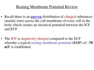
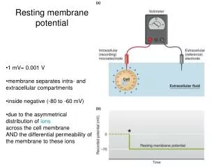
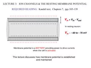
RESTING MEMBRANE POTENTIAL The resting membrane potential is the potential difference that exists across the membrane of excitable cells such as nerve and muscle in the period between action potentials (i.e., at rest). As stated previously, in expressing the membrane potential, it is conventional to refer the intracellular potential to the extracellular potential. The resting membrane potential is established by diffusion potentials, which result from the concentration differences for various ions across the cell membrane. Each permeant ion attempts to drive the membrane potential toward its own equilibrium potential. Ions with the highest permeabilities or conductances at rest will make the greatest contributions to the resting membrane potential, and those with the lowest permeabilities will make little or no contribution. The resting membrane potential of excitable cells falls in the range of −70 to −80 mV. These values can be explained by the concept of relative permeabilities of the cell membrane. Thus, the resting membrane potential is close to the equilibrium potentials for K+ and Cl− because the permeability to these ions at rest is high. The resting membrane potential is far from the equilibrium potentials for Na+ and Ca2+ because the permeability to these ions at rest is low
Action Potentials The action potential is a phenomenon of excitable cells, such as nerve and muscle, and consists of a rapid depolarization (upstroke) followed by repolarization of the membrane potential. Action potentials are the basic mechanism for transmission of information in the nervous system and in all types of muscle. Depolarization is the process of making the membrane potential less negative. As noted, the usual resting membrane potential of excitable cells is oriented with the cell interior negative. Depolarization makes the interior of the cell less negative, or it may even cause the cell interior to become positive. A change in membrane potential should not be described as “increasing” or “decreasing” because those terms are ambiguous. (For example, when the membrane potential depolarizes, or becomes less negative, has the membrane potential increased or decreased?) ♦ Hyperpolarization is the process of making the membrane potential more negative. As with depolarization, the terms “increasing” or “decreasing” should not be used to describe a change that makes the membrane potential more negative.
CHARACTERISTICS OF ACTION POTENTIALS Action potentials have three basic characteristics: stereotypical size and shape, propagation, and all-or-none response. Stereotypical size and shape. Each normal action potential for a given cell type looks identical, depolarizes to the same potential, and repolarizes back to the same resting potential. Propagation. An action potential at one site causes depolarization at adjacent sites, bringing those adjacent sites to threshold. Propagation of action potentials from one site to the next is nondecremental. All-or-none response. An action potential either occurs or does not occur. If an excitable cell is depolarized to threshold in a normal manner, then the occurrence of an action potential is inevitable. On the other hand, if the membrane is not depolarized to threshold, no action potential can occur. Indeed, if the stimulus is applied during the refractory period, then either no action potential occurs, or the action potential will occur but not have the stereotypical size and shape.
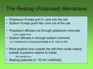
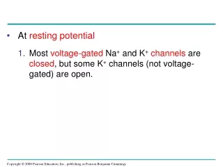
Ionic Basis of the Action Potential The action potential is a fast depolarization (the upstroke), followed by repolarization back to the resting membrane potential. The following Figure illustrates the events of the action potential in nerve and skeletal muscle, which occur in the following steps: 1. Resting membrane potential. At rest, the membrane potential is approximately −70 mV (cell interior negative). The K+ conductance or permeability is high and K+ channels are almost fully open, allowing K+ ions to diffuse out of the cell down the existing concentration gradient. This diffusion creates a K+ diffusion potential, which drives the membrane potential toward the K+ equilibrium potential. The conductance to Cl− (not shown) also is high, and, at rest, Cl− also is near electrochemical equilibrium. At rest, the Na+ conductance is low, and, thus, the resting membrane potential is far from the Na+ equilibrium potential.
2. Upstroke of the action potential. An inward current, usually the result of current spread from action potentials at neighboring sites, causes depolarization of the nerve cell membrane to threshold, which occurs at approximately −60 mV. This initial depolarization causes rapid opening of the activation gates of the Na+ channel, and the Na+ conductance promptly increases and becomes even higher than the K+ conductance 3. Repolarization of the action potential. The upstroke is terminated, and the membrane potential repolarizes to the resting level as a result of two events. First, the inactivation gates on the Na+ channels respond to depolarization by closing, but their response is slower than the opening of the activation gates. Thus, after a delay, the inactivation gates close the Na+ channels, terminating the upstroke. Second, depolarization opens K+ channels and increases K+ conductance to a value even higher than occurs at rest 4. Hyperpolarizing afterpotential (undershoot). For a brief period following repolarization, the K+ conductance is higher than at rest and the membrane potential is driven even closer to the K+ equilibrium potential (hyperpolarizing afterpotential). Eventually, the K+ conductance returns to the resting level, and the membrane potential depolarizes slightly, back to the resting membrane potential. The membrane is now ready, if stimulated, to generate another action potential.
The Refractory Period Absolute refractory period is the period during which another action potential cannot be elicited, Explanation: The inactivation gates of the Na + channel are closed and will remain closed until repolarization occurs. No action potential can occur until the inactivation gates open. Relative refractory period -begins at the end of the absolute refractory period and continues until the membrane potential returns to the resting level. -An action potential can be elicited during this period if a stronger than usual current is provided. Explanation:The slow return of the K channels to the closed state explains the after-hyperpolarization and hence relative refractory period During this time, only a very strong depolarization can overcome the repolarization effects of the open K+ channels and produce a second action potential
Propagation of Action Potentials Propagation of action potentials down a nerve or muscle fiber occurs by the spread of local currents from active regions to adjacent inactive regions. . At rest, the entire nerve axon is at the resting membrane potential, with the cell interior negative. Action potentials are initiated in the initial segment of the axon, nearest the nerve cell body. Spread of depolarization down a nerve fiber by local currents. A, The initial segment of the axon has fired an action potential, and the potential difference across the cell membrane has reversed to become inside positive. The adjacent area is inactive and remains at the resting membrane potential, inside negative. B, At the active site, positive charges inside the nerve flow to the adjacent inactive area. C, Local current flow causes the adjacent area to be depolarized to threshold and to fire action potentials; the original active region has repolarized back to the resting membrane potential.
If the entire nerve were coated with the lipid myelin sheath, however, no action potentials could occur because there would be no low resistance breaks in the membrane across which depolarizing current could flow. Therefore, it is important to note that at intervals of 1 to 2 mm, there are breaks in the myelin sheath, at the nodes of Ranvier. At the nodes, membrane resistance is low, current can flow across the membrane, and action potentials can occur. Thus, conduction of action potentials is faster in myelinated nerves than in unmyelinated nerves because action potentials “jump” long distances from one node to the next, a process called saltatory conduction.