Resting Membrane Potential
Resting Membrane Potential. Esraa kiwan. Membrane Potential. Membrane potential: separation of opposite charges across the plasma membrane difference in the relative number of cations (+ ve ) & anions (- ve ) in the ICF & ECF


Resting Membrane Potential
E N D
Presentation Transcript
Resting Membrane Potential Esraa kiwan
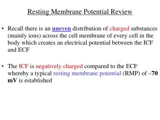
Membrane Potential Membrane potential: separation of opposite charges across the plasma membrane difference in the relative number of cations (+ve) & anions (-ve) in the ICF & ECF These separation produce energy differences between inside and outside which called potential This energy is electrical energy and is measured by Millivolts
Membrane Potential No membrane potentialAn equal number of (+ve) and (-ve) charges are on each side of the membrane Membrane potential exist Unequal number of (+ve) and (-ve) charges are on each side of the membrane
Membrane Potential Separated charges accumulated in a thin layer along the outer and inner surface of the plasma membrane
The magnitude of the potential depends on the degree of separation of the opposite charges The greater the number of separated charges the larger potential Membrane Potential
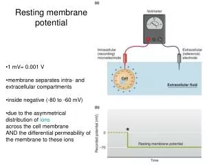
Resting Membrane Potential All plasma membrane of all living cells have resting membrane potential: Varies in value according to the type of the cell: Nerve cell -70 mV Skeletal and cardiac muscle -80 to -90 mV Smooth muscle -60mV The sign always represent the polarity of the excess charge on the inside of the membrane Inside is more -vethan outside
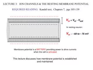
Resting Membrane Potential In the body, the electrical charges are carried by ions The ions primarily responsible for the generation of resting membrane potential are: • Na + • K + • A - (-vecharged intracellular proteins)
The electrical prosperities of the cell membrane are due to: Unequal distribution of a key ions across the plasma membrane Selective movement of these ions through plasma membrane Resting Membrane Potential
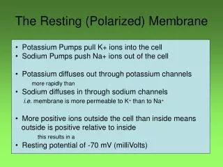
Resting membrane potential Unequal distribution of a key ions between ICF and ECF are maintained by Na+ /K + pump: • Active transport mechanism • Pumps 3Na+ outside, 2K+ inside • Maintains high [K+] inside the cell, high [Na+] outside • Unequal transport generates a membrane potential • Outside becoming more +ve than inside (more +ve are transported out than in)
Resting membrane potential 80% of resting membrane potential is caused by the passive diffusion of K+and Na +down concentration gradient Na +& K+concentration gradient maintained by Na + /K+pump K+permeability about 50-75 X than for Na + K+ions influence the resting membrane potential than Na + Na + / K +pump responsible of 20% of resting membrane potential through maintaing concentration gradient for Na +, K+
Resting Membrane Potential Electrochemical gradient for Na+, K+ in resting state: Concentration gradient: Na + inside K + outside Electrical gradient ( toward the negative charge) Na + inside K + inside
Resting Membrane Potential K+equilibrium potential (EK+):
Resting Membrane Potential Na +equilibrium potential (ENa+):
Resting Membrane Potential Effect of both K+and Na +on resting membrane potential: Na +equilibrium potential (ENa+)= +60 mV K+equilibrium potential (EK+)= -90 mV K+exerts the dominant effect on the resting membrane potential, because the membrane is more permeable to K+resting membrane potential is much closer to EK+than to ENa+ which is -70mV
Changes in Resting Membrane Potential Excitable cell (Nerve & muscle) able to undergo transient, repaid changes in their resting membrane potential These changes serve as electrical signal: Nerve cell: use electrical signal to receive, process, initiate, and transmit messages Muscle: these electrical signals initiate contraction
Changes in Resting Membrane Potential • Polarization: resting state (-ve inside compared with outside) • Depolarization: decrease the negativity of the membrane ( moving toward 0 mV) • Repolarization: the membrane returns to resting potential • Hyperpolarization: the membrane become more negative than at rest (increase negativity)
Changes in Resting Membrane Potential Changes in ions movement a cross the membrane (changes in membrane permeability): If net inward of +ve increases compared to the resting state, the membrane become depolarize (decrease -ve inside) If net outward of +ve increases compared to the resting state, the membrane become hyperpolarized (increase -ve inside)
Changes in resting membrane potential Changes in membrane potential could be: Graded potential Action potential
Action Potential Brief, repaid, large changes in membrane potential The potential actually reverses the inside of the cell become more +ve than outside
Action Potential Stages of action potential Triggering event causes the membrane to depolarize from resting potential (-70 mV) Depolarization proceeds slowly at first, until it reaches a critical level, (threshold potential) (-50 _ -55 mV) At threshold potential, an explosive depolarization takes place: Upward deflection to +30mV Reversal of polarity( the inside of the cell becomes more +ve than outside) Then, the membrane repolarizes, dropping back to resting potential
Action Potential Action Potential is caused by marked changes in membrane permeability of ions: At rest, membrane permeability to K+is much higher than it for Na + So, K+makes the greatest contribution to the resting membrane potential. This will completely change during action potential, due to activation of Voltage-GatedNa +&K+Channels
Action Potential Voltage Gated Channels • The channels that their state (opening or closure) depends on the voltage changes of the membrane • At resting potential, all the Voltage-Gated Channels are closed
Action potential When a membrane starts to depolarize toward threshold, some of the voltage-gated Na+channels will open Then, Na+will move down its electrochemical gradient into the cell This influx of Na+will depolarize the membrane more, & so more channels will open… & so on
Action potential At threshold, there is an explosive increase in permeability of Na+ (≈600 times more permeable to Na+than to K+) The membrane potential will become closer to ENa+= +60 mV The potential will be (+30 mV) Before reaching the Peak-Potential, Na+channels will be inactivated, so no more Na+ leak to the inside
Action potential Simultaneous with inactivation of Na+channels, the voltage-gated K+channels starts to slowly open, with maximum opening occurring at the peak of action potential These channels will greatly increase the K+permeability (≈300 times more than Na+) This will lead to marked efflux of K+out of the cell, down its concentration gradient This will rapidly restore the negative resting potential
Action potential Rising phase: from threshold to (+30 mV) is caused by Na+influx Falling phase: from (+30 mV) to resting potential is caused by K+efflux Ions distribution is restored by the action of Na+-K+ pump
Action potential Action Potential is an all-or-none response • If the membrane is not depolarized to its threshold level, no action potential is produced • If the stimulus strength is increased more than what is needed to produce action potential there will be no change in magnitude of action potential (produce same action potential) • Strong stimulus produces more action potential (increase in #), and cause more cells to reach their threshold
Action potential Propagation of action potential throughout nerve fiber • Once action potential is generated in any part of neuron, it will propagated all the way through the axon to reach axon terminal • Action potential spreads throughout the membrane in undecresing fashion (non-decremental)
Action potential Contiguous conduction in unmyelinated fibers: Action potential at one point will excited the adjacent point by increasing the permeability of adjacent point to Na+ causing generation of Action Potential Propagation of action potential throughout nerve fiber
Action potential Saltatory conduction in myelinated fibers Propagation of action potential throughout nerve fiber
Refractory Period • A period during which a new action potential cannot be initiated by normal events in a region that has just undergone an action potential • Two phases: • Absolute Phase: a second action potential can’t be produced, no matter how strong is the stimulus. The membrane completely unresponsive to second stimulus at all • Relative Phase: during which the second action potential can be produced only by an exceptionally strong triggering
Prosperities of Action Potential • Is an “all or none” event (either pass threshold or dosen’t) • Is propagated down the axon in nondecremental fashion • Has a fixed amplitude ( action potential do NOT change in Height) • Has a refractory period (in which stimulation will not produce an action potential)
Graded Potential • Is a local change in the membrane potential that occurs in varying grades (changes may be 10 or 20 mV) • The magnitude vary with the strength of the stimulus, the stronger the triggering event the larger the graded potential • The magnitude of the graded potential continues to decrease the farther it moves away from the initial active area. (Decremental) • Graded potential do not have a refractory period
A neuron may terminate at: • A muscle • A gland • Another neuron
Synaptic Transmission • A synapse is the junction between two neurons • It involves the junction between an axon terminal of one neuron (presynaptic neuron), & the dendrites or the cell body of a second neuron (postsynaptic neuron)
Synaptic Transmission • The action potential (the electrical signal) crosses the synapse by chemical means to induce electrical changes in other neuron • The chemicals released from one neuron to induce electrical changes in next neuron are neurotransmitters • Acetylcholine • Norepinephrine • Histamine • Glycine • Gamma-aminobutyric acid (GABA) • Synapses operate in one directiononly
Synaptic Transmission • Events taking place in a synapse: • Propagation of an action potential to the axon terminal of a presynaptic neuron • The arriving action potential produces an influx of calcium ionsthrough voltage-dependent calcium ion channels. ([Ca+2] in ECF is much higher than that inside the synaptic knob) • Ca+2 induces the release by exocytosis of neurotransmitters from synaptic vesicles into the synaptic cleft • After diffusing across the cleft, the neurotransmitter binds with its receptor sites on the subsynaptic membrane • This binding triggers the opening of specific ion channels in the subsynaptic membrane. This alters the permeability of the postsynaptic neuron (Chemical messenger-gated channels)
Synaptic Transmission • The changes in postsynaptic neuron by neurotransmitter could be • excitatoryor inhibitory
Types of synapses • Excitatory synapse: • the response to the receptor-neurotransmitter combination is an opening of Na+& K +channels within the subsynaptic membrane. • Na+moves into the postsynaptic neuron & K +out of it, but to a less extent • The result is a net movement of positive ions into the cell Small depolarizationin the postsynaptic neuron • Activation of one excitatory synapse can rarely depolarize the postsynaptic membrane sufficiently to bring it to threshold, but it brings it closer to threshold • This postsynaptic potential change is called: Excitatory PostSynaptic Potential (EPSP)
Types of synapses • Inhibitory synapse: • The response to the receptor-neurotransmitter combination is an opening of K+& Cl -channels within the subsynaptic membrane • The resulting ion movements bring about a small hyperpolarizationof the postsynaptic neuron (greater internal negativity) • This moves the membrane potential away from threshold • This small hyperpolarization of the postsynaptic cell is called: Inhibitory PostSynaptic Potential (IPSP)
Synaptic Transmission • The time needed for action potential to cause the release of the neurotransmitter into synaptic cleft and produce its effect on postsynaptic neuron is called synaptic delay, its about 0.5-1 msec
Synaptic Transmission • Fate of neurotransmitter that released from presynaptic membrane: • Uptake of the neurotransmitter into the cytoplasm of the presynaptic membrane (reused again) • Enzymatic deactivation: inactivation of neurotransmitter by certain enzyme
Synaptic Transmission • Grand PostSynaptic Potential (GPSP): summation of all potentials ( both EPSP + IPSP) that occur at the same time • The postsynaptic neuron can be brought to threshold by: • Temporal summation: is the summing of several EPSPs occurring very close together in time because of successive firing of a single presynaptic neuron • Spatial summation: is the summation of EPSPs originating simultaneously from several different presynaptic inputs (from different points in space) • If an excitatory & an inhibitory input are simultaneously activated, the concurrent EPSP & IPSP more or less cancel each other out.