Download
1 / 34
350 likes | 385 Vues
Explore the mechanisms of gas exchange in animals, from respiratory surfaces to specialized body parts like gills and lungs. Discover the adaptations that allow animals to thrive in challenging environments such as high altitudes. Learn how efficient gas exchange is vital for energy metabolism and overall survival.

E N D
Surviving in Thin Air • The air at the height of the world’s highest peak, Mt. Everest, is very low in oxygen • Even expert mountain climbers do not always survive the journey • Thin air can weaken muscles, damage the digestive system, cloud the mind, and sometimes fill the lungs with blood
Their efficient lungs draw more oxygen from the atmosphere • Their hemoglobin has a high affinity for oxygen • They have a large number of capillaries to deliver this oxygen-rich blood to tissues and muscles • Geese have adaptations that allow them to fly over the Himalayas
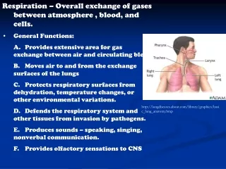
MECHANISMS OF GAS EXCHANGE • Gas exchange is the interchange of O2 and CO2 between an organism and its environment • It is also called respiration
22.1 Overview: Gas exchange involves breathing, the transport of gases, and the servicing of tissue cells • Gas exchange is essential because energy metabolism requires O2 and produces CO2 • There are three phases of gas exchange
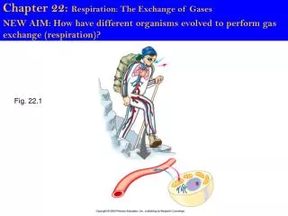
O2 Lung CO2 1 Breathing Circulatorysystem 2 Transportof gases bythe circulatorysystem Mitochondria 3 O2 Servicing ofcells withinthe bodytissues CO2 Capillary Cell Figure 22.1
22.2 Animals exchange O2 and CO2 through moist body surfaces • O2 enters an animal and CO2 leaves by diffusion through a respiratory surface • Respiratory surfaces are made up of living cells
Example: earthworms • Some animals use their entire skin as a gas-exchange organ Cut Cross sectionof respiratorysurface (theskin coveringthe body) CO2 O2 Capillaries Figure 22.2A
Gills in fish • In most animals, specialized body parts carry out gas exchange Body surface Respiratorysurface(gill) Capillaries CO2 O2 Figure 22.2B
Lungs in land vertebrates • Tracheae in insects Body surface Body surface Respiratorysurface(tracheae) Respiratorysurface(within lung) O2 Body cells(no capillaries) CO2 O2 Capillary CO2 Figure 22.2C, D
22.3 Gills are adapted for gas exchange in aquatic environments • Gills are extensions of the body that absorb O2 dissolved in water • In fish, gill filaments bear numerous platelike lamellae • Lamellae are packed with blood vessels • They are the respiratory surfaces
Gill arch Directionof waterflow • The structure of fish gills Gill arch Bloodvessels Gillfilaments Oxygen-poorblood Oxygen-richblood Lamella Waterflow Figure 22.3
22.4 Countercurrent flow in the gills enhances O2 transfer • Blood flows through the lamellae in a direction opposite to water flow • This countercurrent maintains a diffusion gradient that maximizes the uptake of O2 Water flowover lamellae Blood flowthroughlamellae Figure 22.4
22.5 The tracheal system of insects provides direct exchange between the air and body cells • Land animals exchange gases by breathing air • Air contains more O2 and is easier to move than water • But water loss from the respiratory surfaces can be a problem
O2 diffuses from the finely branched tubes directly into cells • In insects, a network of tracheal tubes carries out gas exchange Figure 22.5B
Air sacs Tracheae Openingfor air Bodycell Tracheole Airsac Trachea Air Body wall Figure 22.5A, C
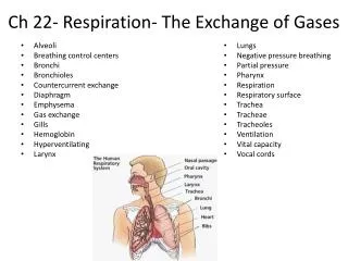
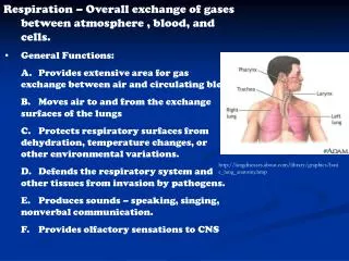
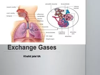
22.6 Terrestrial vertebrates have lungs • In humans and other mammals, air enters through the nasal cavity • It passes through the pharynx and larynx into the trachea • The trachea forks to form two bronchi • Each bronchus branches into numerous bronchioles
The human respiratory system Nasalcavity Pharynx (Esophagus) Left lung Larynx Trachea Rightlung Bronchus Bronchiole Diaphragm (Heart) Figure 22.6A
Alveoli form the respiratory surface of the lungs • Oxygen diffuses through the thin walls of the alveoli into the blood • The bronchioles end in clusters of tiny sacs called alveoli Figure 22.6C Oxygen-richblood Oxygen-poorblood Bronchiole Alveoli Blood capillaries Figure 22.6B
22.7 Connection: Smoking is one of the deadliest assaults on our respiratory system • Mucus and cilia in the respiratory passages protect the lungs • Pollutants, including tobacco smoke, can destroy these protections • Smoking kills about 430,000 Americans each year
Smoking also causes emphysema • Cigarette smoke makes alveoli brittle, causing them to rupture • This reduces thelungs’ capacity for gas exchange • Smoking causes lung cancer and contributes to heart disease Figure 22.7A, B
22.8 Breathing ventilates the lungs • Breathing is the alternation of inhalation and exhalation Rib cageexpands asrib musclescontract Rib cagegets smalleras rib musclesrelax Airinhaled Airexhaled Lung Diaphragm INHALATIONDiaphragm contracts(moves down) EXHALATIONDiaphragm relaxes(moves up) Figure 22.8A
But our lungs hold more than this amount • The alveoli do not completely collapse • A residual volume of “dead” air remains in the lungs after exhalation • Vital capacity is the maximum volume of air we can inhale and exhale
These structures act as bellows that keep air flowing through the lungs • However, they do not function directly in gas exchange • Other organisms, such as birds, have air sacs Air Air Anteriorair sacs Trachea Posteriorair sacs Lungs Lungs Airtubesin lung 1 mn INHALATION:Air sacs fill EXHALATION:Air sacs empty; lungs fill Figure 22.8B
22.9 Breathing is automatically controlled • Breathing control centers are located in the pons and medulla of the brain • These automatic controls keep breathing in tune with body needs
This triggers a cascade of events Brain Cerebrospinal fluid • During exercise, the CO2 level in the blood rises, lowering the blood pH BREATHING CONTROLCENTERS—stimulated by: Pons Medulla CO2 increase / pH decreasein blood Nerve signalindicating lowO2 level Nerve signalstriggercontractionof muscles O2 sensorin artery Diaphragm Figure 22.9 Rib muscles
TRANSPORT OF GASES IN THE BODY 22.10 Blood transports the respiratory gases, with hemoglobin carrying the oxygen • The heart pumps oxygen-poor blood to the lungs • In the lungs it picks up O2 and drops off CO2 • In the tissues, cells pick up CO2 and drop off O2 • Gases diffuse down pressure gradients in the lungs and the tissues
Gas exchange in the body Figure 22.10A
It carries most of the oxygen in the blood • Hemoglobin is a protein in red blood cells Hemegroup Iron atom O2 loadedin lungs O2 O2 unloadedin tissues O2 Polypeptide chain Figure 22.10B
22.11 Hemoglobin helps transport CO2 and buffer the blood • Hemoglobin helps buffer the pH of blood and carries some CO2
TISSUE CELL CO2 produced INTERSTITIALFLUID CO2 • Most CO2 in the blood combines with water to form carbonic acid • The carbonic acid breaks down to form H+ ions and bicarbonate ions • These help buffer the blood BLOODPLASMAWITHINCAPILLARY CO2 Capillarywall CO2 H2O REDBLOODCELL Hemoglobinpicks upCO2 and H+ H2CO3 Carbonic acid HCO3– + H+ Bicarbonate HCO3– Figure 22.11A
ALVEOLAR SPACE IN LUNG CO2 CO2 • Most CO2 is transported to the lungs in the form of bicarbonate ions CO2 CO2 H2O HemoglobinreleasesCO2 and H+ H2CO3 HCO3– + H+ HCO3– Figure 22.11B
22.12 Connection: The human fetus exchanges gases with the mother’s bloodstream Placenta, containingmaternal blood vesselsand fetal capillaries • A human fetus depends on the placenta for gas exchange Umbilical cord,containing fetalblood vessels Amnioticfluid Uterus Figure 22.12
A network of capillaries exchanges O2 and CO2 with maternal blood that carries gases to and from the mother’s lungs • At birth, increasing CO2 in the fetal blood stimulates the fetus’s breathing control centers to initiate breathing