Download
1 / 17
190 likes | 455 Vues
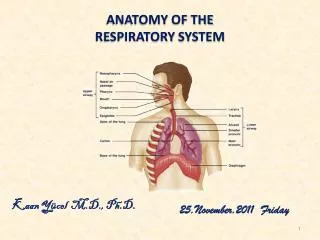
Functional Anatomy of the Respiratory System. Dr. Meg- angela Christi Amores. Pulmonary Ventilation. Pulmonary Ventilation – inflow and outflow of air between the atmosphere and the lungs Muscles for Respiration: Diaphragm External Intercostal muscles Sternocleidomastoid Muscles

E N D
Functional Anatomy of the Respiratory System Dr. Meg-angela Christi Amores
Pulmonary Ventilation • Pulmonary Ventilation – inflow and outflow of air between the atmosphere and the lungs • Muscles for Respiration: • Diaphragm • External Intercostal muscles • Sternocleidomastoid Muscles • Anterior Serrati • Scalene muscles • Abdominal Rectus musles • Internal Intercostals
Lung Expansion and Contraction 2 ways: • Diaphragm Movement • or – lengthen or shorten chest cavity • Ribs • Elevate or depress – increase or decrease antero-posterior diameter of chest cavity Normal quite breathing is accomplished almost entirely by first method.
Diaphragm Movement • During INSPIRATION: • Diaphragm contracts and pulls lower surface of the lung downward • During EXPIRATION: • Diaphragm relaxes accompanied by elastic recoil of lungs, chest wall and abdominal structures During heavy breathing, extra force is achieved mainly by contraction of abdominal muscles
Ribs Movement • During INSPIRATION • Ribs project almost entirely forward from an original downward position • Sternum also moves forward away from spine • Anteroposterior (AP) diameter increases to 20% • Muscles that elevate ribs: • External intercostals • Sternocleidomastoid • Anterior Serratus • Scalene Muscles
PRESSURES • Lungs are “elastic” – collapses like a balloon when there is no force to keep it inflated • There are no attachments between the lungs and the ribcage except at hilum • Lungs float in pleural fluid • Lymphatics provide slight suction between visceral surface of lung pleura and parietal surface of thoracic cavity
Pleural Presure • Pressure of fluid in the narrow space between lung pleura and chest wall pleura • Slightly negative pressure • At beginning of inspiration: -5 cmH20 • The amount needed to hold the lungs open • During inspiration: -7.5cmH20 As negativity increases, lung volume increases to 0.5L
Alveolar Pressure • Pressure of air inside the lung alveoli • Open glottis – pressures are equal at 2 atm • For inspiration – inward flow of air into alveoli the pressure must fall to a value slightly below atmospheric pressure (below 0) • During inspiration: alv pressure drops to -1cmH20 = 0.5 L of air
Compliance • Compliance is the extent to which lungs expand for each unit of increase in transpulmonary pressure • = 200mL/ 1 cmH20 change in transpulmonary pressure
Work of breathing • Equivalent to Work of Inspiration • 3 fractions: • That required to expand the lungs against the lung and chest elastic forces = compliance work • That required to overcome the viscosity of the lung and chest wall structures =tissue resistance work • The required to overcome airway resistance during the movement of air into the lungs = airway resistance work
Pulmonary volumes and capacities • Spirometry – process of studying pulmonary ventilation, recording the volume movement of air into and out of lungs • Pulmonary Volumes: • Tidal Volume: vol. of air inspired/expired with each normal breathing = 500 mL • Inspiratory Reserve Volume – maximum extra volume of air that can be inspired over and above normal tidal volume = 300 mL
Pulmonary volumes and capacities 3. Expiratory Reserve Volume : maximum extra volume of air that can be expired forcefully after end of a normal tidal expiration = 1.1L • Residual Volume : volume of air remaining in the lungs after most forceful expiration = 1.2L
Pulmonary volumes and capacities • Pulmonary Capacities • Two or more volumes togethere • Inspiratory Capacity : TV + IRV = 3.5L • Functional Residual Capacity: ERV+RV = 2.3L • Vital Capacity : IRV + TV + ERV = 4.6L • Total Lung Capacity: VC + RV = 5.8L All pulmonary volumes and capacities are about 20-25% less in women than in men.
For the next meeting, read on Pulmonary Gas exchange and Gas transport • Guyton Textbook of Medical Physiology