Download
1 / 28
280 likes | 548 Vues
Exercise 26 Functional Anatomy of The Urinary System. Gross Anatomy of the Human Urinary System. Renal capsule a tough fibrous layer surrounding the kidney and covered in a thick layer of adipose tissue. It provides some protection from trauma and damage . Organs of the Urinary System.

E N D
Gross Anatomy of the Human Urinary System Renal capsule a tough fibrous layer surrounding the kidney and covered in a thick layer of adipose tissue. It provides some protection from trauma and damage.
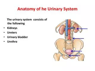
Organs of the Urinary System • Kidneys • Ureters • Urinary bladder • Urethra • Renal Artery Figure 15.1a
Internal Anatomy of the Kidney Renal cortex —outer region; contains most of nephron structure Renal medulla —inside the cortex; contains inner collecting ducts Renal pelvis – basin continuous with ureter Renal columns —extensions of cortex-like material inward that separate the pyramids Calyces —cup-shaped structures, extensions of pelvis that funnel urine Renal or fibrous capsule -- a tough fibrous layer surrounding the kidney
Functional Microscopic Anatomy of the Kidney m k l n o b a c Interlobular vein h e d f Interlobular artery g i j
n c a g e b h f d k
Glomerulus= site of filtrate formation Red arrows = Blood Blue arrows = Filtrate Glomerulus = a tiny ball-shaped structure composed of capillary blood vessels actively involved in the filtration of the blood to form urine . Bowman’s Capsule =Fluids from blood in the glomerulus are collected in the Bowman's capsule (i.e., glomerular filtrate) and further processed along the nephron to form urine.
Proximal convoluted tubule= primary site of tubular reabsorption Glucose, Amino Acids, Na+, and Water are being reabsorbed by the Proximal Convoluted Tubule
Collecting Ducts- collects the urine from the nephron and send it to the ureter.
Peritubular capillaries – receives substances from the tubular cells
Bowman’s Capsule: the inner membrane forms part of the filtration mechanism • There are two layers of cells that fluid (with its contents) has to pass through during filtration. The second layer is the visceral layer of the Bowman’s capsule, formed by podocytes The first layer is the endothelium of the glomerular capillary.
The renal corpuscle is where plasma is filtered from capillaries into the renal tubules. At the center of renal corpuscle is the glomerulus, a meshwork of capillaries. The glomerulus is surrounded by Bowman's capsule
Glomerulus and Bowman’s Capsule Visceral layer Parietal layer Podocytes make up the visceral layer of Bowman’s Capsule
Glomerulus is a high-pressure capillary bed The afferent arteriole feeding the glomerulus is is larger than the efferent arteriole which drains the arteriole The high pressure forces out fluid and blood components smaller than proteins from the glomerulus into the glomerular capsule
Proximal Convoluted Tubule The cuboidal cells of the proximal convoluted tubule have long microvilli (brush border) on their inside surface that dramatically increase the surface area for reabsorption from the filtrate. Filtrate What is the purpose of this brush border epithelium? Close up of brush border (microvilli)
n c a g e b h f d k
Glomerular capsule Proximal convoluted tubule Loop of Henle Distal convoluted tubule Collecting duct Nephron in Cortex Renal Cortex Medullary pyramid Calyces Renal pelvis
Kidney This is a low power view of a cross section through the kidney. Note the inner medullary tissue (green) surrounded by the outer cortical tissue (blue)
Cortex Identify a glomerulus, which appears as a ball of tightly packed material containing many small nuclei Notice the vacant appearing region corresponding to the glomerular capsule that surrounds it. The balance of the kidney tissue consists of renal tubules.
Loop of Henle (D) Descending limbs of the loop of Henle look similar to the proximal tubule, with apical brush borders. (Blue) (A) Ascending limbs are composed of cuboidal cells, but unlike the proximal convoluted tubule, they do not have apical brush borders. (Green) (C) Collecting ducts can also be seen on this slide. They can be easily distinguished by the presence of prominent lateral borders between adjacent cells. (Orange)
Loop of Henle • Descending limb, with its brush border similar to that of the proximal tubule. (Blue) • Ascending limb lacks this brush border and its cells have a more squamous appearance. (Red) • Collecting duct can be observed. (Purple)
Renal Pelvis Urine in the collecting ducts eventually empties into the renal pelvis. Renal pelvis is the initial dilated portion of ureter.
Urinary Bladder The urinary bladder is lined by transitional epithelium, underneath which are thick layers of smooth muscle interwoven in various directions. Compare the transitional epithelium between a relaxed bladder and a distended bladder.