Download
1 / 53
720 likes | 1.13k Vues
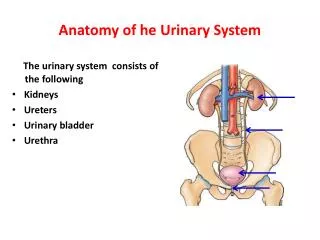
PAN 201. Anatomy of Urinary System. URINARY SYSTEM ORGANS. Kidneys (2) Ureters (2) Urinary bladder Urethra. KIDNEY FUNCTIONS. Control blood volume and composition. KIDNEY FUNCTIONS. Filter blood plasma, eliminate wastes Regulate blood volume, pressure Regulate fluid osmolarity

E N D
PAN 201 Anatomy of Urinary System
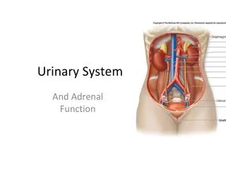
URINARY SYSTEM ORGANS • Kidneys (2) • Ureters (2) • Urinary bladder • Urethra
KIDNEY FUNCTIONS • Control blood volume and composition
KIDNEY FUNCTIONS • Filter blood plasma, eliminate wastes • Regulate blood volume, pressure • Regulate fluid osmolarity • Secrete renin • Secrete erythropoietin (EPO) • Regulate PCO2, Acid-Base balance • Synthesize calcitriol (Vitamin D) • Detoxify free radicals, drugs • Gluconeogenesis
EXCRETION Removal of wastes • Respiratory system • CO2, water • Integumentary system • Water, salts, lactic acid, urea • Digestive system • Water, salts, CO2, lipids, bile pigments, cholesterol, etc. • Urinary system • Metabolic wastes, toxins, drugs, hormones, salts, H+, water
KIDNEY ANATOMY Protected by three connective tissue layers • Renal fascia • Attaches to abdominal wall • Adipose capsule • Fat cushioning kidney • Renal capsule • Fibrous sac • Protects from trauma and infection
KIDNEY ANATOMY Gross anatomy • Renal sinus • Renal parenchyma
KIDNEY ANATOMY Renal sinus • Surrounded by renal parenchyma • Contains blood & lymph vessels, nerves, urine-collecting structures
KIDNEY ANATOMY Renal parenchyma • Glandular tissue • Forms urine • Two zones • Outer cortex • Inner medulla
KIDNEY ANATOMY Renal parenchyma • Renal pyramids • Extensions of cortex (renal columns) divide medulla into 6 – 10 renal pyramids • Pyramid + overlying cortex = Lobe • Point of pyramid = Papilla • Papilla nested in cup (minor calyx) • 2 – 3 minor calices Major calyx • 2 – 3 major calices Renal pelvis • Renal pelvis Ureter
KIDNEY ANATOMY: NEPHRONS Nephrons • Functional units of kidney • ~1.2 million per kidney • Three main parts • Blood vessels • Renal corpuscle • Renal tubule
NEPHRONS Blood vessels servicing kidney • Supplied by renal artery • ~21% or cardiac output • (Mass in only ~ 0.4%) • Afferent arterioles • Capillary cluster (glomerulus)
NEPHRONS Blood vessels servicing kidney • Glomerulus • Fenestrated capillaries • Capillary filtration in glomerulus initiates urine production • Filtrate lacks cells & proteins • Drained by efferent arteriole • Peritubular capillaries • Renal vein
NEPHRONS Renal corpuscle • Glomerulus plus capsule • Glomerulus enclosed in two-layered glomerular capsule • “Bowman’s capsule” • Fluid filters from glomerular capillaries • “Glomerular filtrate” • Fluid collects in capsular space • Fluid flows into renal tubule
NEPHRONS Renal tubule • Leads from glomerular capsule • Ends at tip of medullary pyramid • ~3 cm long • Four major regions • Proximal convoluted tubule • Nephron loop • Distal convoluted tubule • Collecting duct
NEPHRONS Renal tubule • Proximal convoluted tubule (PCT) • Arises from glomerular capsule • Longest, most coiled region • Prominent microvilli • Function in absorption • Much contact with peritubular capillaries
NEPHRONS Renal tubule • Nephron loop (“Loop of Henle”) • “U” – shaped, distal to PCT • Descending and ascending limbs • Thick segments • Active transport of salts • High metabolism, many mitochondria • Thin segments • Permeable to water • Low metabolism
NEPHRONS Renal tubule • Distal convoluted tubule (DCT) • Coiled, distal to nephron loop • Shorter than PCT • Less coiled than PCT • Very few microvilli • Contacts afferent and efferent arterioles (regulation imparted) • Contact with peritubular capillaries
NEPHRONS Renal tubules • Collecting duct • DCTs of several nephrons empty into a collecting duct • Passes into medulla • Several merge into papillary duct (~30 per papilla) • Drain into minor calyx
URINE FORMATION Overview • Blood plasma Urine • Four steps • Glomerular filtration • Tubular reabsorption • Tubular secretion • Water conservation
URINE STORAGE Ureters • Carry urine from kidneys to urinary bladder via peristalsis • Rhythmic contraction of smooth muscle • Enter bladder from below • Pressure from full bladder compresses ureters and prevents backflow
Arterial Supply • Ureter is supplied by multiple arteries throughout its course • From above downward, these are:: • Renal artery • Gonadal artery • Common iliac artery • Internal iliac artery 1 2 3 4
URINE STORAGE Ureters • Small diameter • A 25 – 30 cm long muscular tube transporting urine from kidney to urinary bladder. • Begins as a continuation of renal pelvis. • Easily obstructed or injured by kidney stones (renal calculi)
URINE STORAGE Urinary bladder • Muscular sac • Wrinkles termed rugae • Openings of ureters common site for bladder infection
Urinary Bladder • Located immediately behind the pubic symphysis • Shape and relations vary according to the amount of urine it contains • An empty bladder: • In adults, is entirely a pelvic organ; as it fills, rises up into the hypogastric region. • In young children, it projects above the pelvic inlet
An empty bladder is pyramidal in shape having: • Anapex • Abase (posterior surface) • Asuperiorsurface • Twoinfrolateral surfaces • Aneck
Apex • Directed forward • Lies behind the upper margin of the symphysis pubis • Is connected to umbilicus by the median umbilical ligament (remnant of urachus)
Base or Posterior surface • Triangular in shape • Upper part covered by peritoneum • Lower part related to: • In males: vas deferentia and seminal vesicles • In females: vagina
Female pelvis Male pelvis Superior surface Completely covered by peritoneum. Related to the coils of ileum or sigmoid colon in males and to uterus in females
Infrolateral surfaces: • Related in front to the retro pubic pad of fat & the pubic bones • Posteriorlylie in contact with the obturatorinternus above and levatorani below Retropubic fat • Accommodates distention of bladder • Continuous with anterior abdominal wall. • Rupture of bladder results in escape of urine to anterior abdominal wall
Neck: • Lies inferiorly, and is the most fixed part of the bladder • Is related to lower border of symphysis pubis • In male, rests on the upper surface of prostate. Here, the smooth muscle fibers of the bladder are continuous with those of the prostate • The circular muscle fibers thickened to form the sphincter vesicae
Interior of the Urinary Bladder • Mucous membrane thrown into folds except in the triangular region in the base of bladder, between the openings of the two ureters and the urethra. This region is called the ‘trigone’.Here The mucous membrane is always smooth even when the bladder is empty • Uvula vesicae, a small elevation located just behind the urethral orifice, It is produced by the median lobe of prostate.
Blood & Nerve Supply • The nerves form the vesical nerve plexus that contains: • Sympathetic fibers derived mainly fromL1,2 • Parasympathetic fibers derived from pelvic splanchnic nerves S2,3,4 • Sensory fibers from the bladder are visceral and transmit pain sensation resulting from overdistention Arterial supply: from internal iliac artery Venous drainage: into internal iliac vein Lymphatics:into internal iliac lymph nodes
The normal capacity of bladder is about 300-500ml. • As bladder fills, the superior surface bulges upward into abdominal cavity. • The peritoneal lining is peeled off the lower part of anterior abdominal wall and thebladder comes into direct contact with the anterior abdominal wall
URINE ELIMINATION Urethra • Conveys urine from body • Internal urethral sphincter • Retains urine in bladder • Smooth muscle, involuntary • External urethral sphincter • Provides voluntary control over voiding of urine
URINE ELIMINATION Urethra • 3 – 4 cm long in females • Bound by connective tissue to anterior wall of vagina • Urethral orifice exits body between vaginal orifice and clitoris
URINE ELIMINATION Urethra • ~18 cm long in males • Prostatic urethra • ~2.5 cm long, urinary bladder prostate • Membranous urethra • ~0.5 cm, passes through floor of pelvic cavity • Penile urethra • ~15 cm long, passes through penis