Download
1 / 30
711 likes | 3.44k Vues
Anatomy of he Urinary System. The urinary system consists of the following Kidneys Ureters Urinary bladder Urethra. The Kidney. Bean-shaped organ It lies on the posterior abdominal wall at the side of vertebral column It measures 4x2x1 inches

E N D
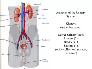
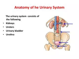
Anatomy of he Urinary System The urinary system consists of the following • Kidneys • Ureters • Urinary bladder • Urethra
The Kidney • Bean-shaped organ • It lies on the posterior abdominal wall at the side of vertebral column • It measures 4x2x1 inches • It has anterior and posterior surfaces and medial and lateral borders • Right kidney is lower than the left because of the liver • The level of kidneys varies with respiration of about 1 inch
The Kidney Cont., Renal hilum • Is found at the medial concave surface • Renal vein, renal artery and ureter are found in the hilum in anterior posterior order • Lymphatics and sympathetic nerves pass through the hilum Renal sinus All structures pass through the hilum in addition to small amount of fat and the pelvis are found in the sinus
Suprarenal glands, kidneys, and ureters and their vessels are retroperitoneal structures • Renal capsule formed of dense connective tissue surrounding the kidneys for support and protection • Perirenal fat a layer of fat surrounds the kidney outside the renal capsule
Kidneys, suprarenal glands, and perirenal fat are enclosed by fascial membrane called Renal fascia • The two layers extend medially to enclose the renal vessels and blend with vascular fascia • The two layers extend inferiorly to enclose the ureters as asPeriuretric fascia • A layer of fat surrounding the kidneys, ureters, suprarenal glands external to renal fascia called Pararenal fat
Perirenal fat, renal fascia, pararenal fat and some collagen fibers hold the kidneys in fixed position • Superiorly, renal fascia is attached to inferior diaphragmatic fascia
Internal structure of the Kidney Each kidney consists of: • Outer cortex • Inner medulla formed of renal pyramids with apex forming renal papilla • Cortex extends into medulla as renal columns • Renal pelvis fills most of the sinus • Major calyces • Minor calyces
Anterior relation of the Kidney Right kidney: • Suprarenal gland • Liver • Duodenum • Right colic flexure • Ileum Left kidney • Suprarenal gland • Stomach • Spleen • Splenic artery • Pancreas • Jejunum • Left colic flexure
Anterior relation of the Kidney Left Kidney Right Kidney
Posterior Relation of the Kidney • Diaphragm separates the kidney from the pleural cavity and 11thand 12th ribs • Psoas major medially • Quadratuslumborum in the middle, transversusabdominis laterally • Subcostal, iliohypogastric and ilioinguinal nerves descend obliquely behind the kidney
The Ureter • It is a muscular organ • Extends from renal pelvis in abdomen, crosses the pelvic brim at common iliac artery bifurcation to the urinary bladder • It 10 inches in length • On the back, extends along a line from a point 5 cm from spine of L1 to posterior superior iliac spine
The Ureter Cont., • The abdominal part is retroperitoneal • It runs close to the tips of transverse processes of lumbar vertebrae as seen in contrast radiographs • It passes vertically on psoas muscle • It enters the pelvis crossing the external iliac artery • It shows three constriction along its course: Between ureter and pelvis At crossing the external iliac artery At entrance to urinary bladder • They are the sites of obstruction by renal calculi
The Ureter Cont., • Ureter runs on the lateral part of lesser pelvis • It runs parallel to medial part of greater sciatic notch • At level of ischial spine, it turns anteromedially to enter the inferior surface of the bladder in inferiomedialdirction • The enterance is 5 cm a part on external surface and 2.5 cm a part at internal surface • Oblique passage creates a sphincter like structure at lower end of ureter
Anterior Relation of the Ureter Duodenum Terminal Ileum Left gonadal vessels Right gonadal vessels Left colic vessels Right colic vessels Sigmoid colon and mesocolon Ileocolic vessels
The Urinary Bladder Body Apex Fundus • It is a hollow muscular organ • It is a pelvic organ after puberty • It lies behind and superior to pubic bones leaving retropubic space in between • It is divided into apex, fundus and body • It has 4 surfaces, superior, posterior (base) and two inferiolateral • It is freely movable except at the neck that is attached by lateral ligaments of bladder and puboprostatic ligament in male andpubovesical ligament in female
The Urinary Bladder Cont., Posterior relation in male: • Vas deferens • Seminal vesicle • Rectum • Rectovesical fascia • Peritoneum Posterior relation in female: • Vagina and part of uterus Superior relation in male: • Peritoneum • Coils of ileum • Sigmoid colon Superior relation in female: • Uterus Lateral relation • Obturatorinternus muscle • Levatorani muscle
The Urinary Bladder Cont., Anterior relation • Symphysis pubis • Retropubic fat Inferior relation: • Prostate gland • The muscle of the bladder wall is called Detrusor muscle • It is thickened at the neck to form involuntary internal urethral sphincter • Trigone is triangular area where the two ureters and urethra open into its angles
Blood Supply of the Urinary System Kidney: • Renal arteries are paired branches arise from aorta at the level of intervertebral disc between L1,2 • Right one longer than the left, passes behind IVC • At the hilum, each one divides into 5 segmental arteries each supplies a renal segment • Renal segments are independent in their blood supply • Blood is drained by segmental veins to renal veins
Blood Supply of the Urinary System Cont., Abdominal part of ureter: • Uretric branches from the renal artery are constant ones. • Other branches from gonadal, aorta and common iliac arteries • They give ascending and descending branches that anastomose with each other • Veins are drained by renal and gonadal veins
Blood Supply of the Urinary System Cont., Pelvic part of ureter • Uretric branches of common iliac, internal iliac and gonadal arteries • Constant branches come from inferior vesical and (in female) uterine arteries • Veins are corresponded to arteries
Blood Supply of the Urinary System Cont., Urinary bladder • Superior vesical artery • Inferior vesical artery • Vaginal artery replace inferior vesiacal artery in female • In male, venous plexus around the bladder and prostate drain into inferior vesical vein • Also, superior vesical vein drains the bladder • Both veins drain into internal iliac vein • In female, venous plexus around bladder drain into vaginal or uterovaginal vein and then to internal iliac vein • Also, superior vesicalvein drains the bladder
Lymphatic drainage Kidney • Into left and right lumbar (aortic and caval) lymph nodes Upper ureter • To kidney lymphatics or to lumbar lymph nodes Middle ureter • To common iliac lymph nodes Lower ureter • To common , external, and internal iliac lymph nodes Pelvic ureter and bladder • To internal iliac lymph nodes
Nerve Supply • Parasympathetic fibers leads to contraction of smooth fibers in ureter and bladder and relaxation of smooth fibers in internal urethric sphincter • Sympathetic fibers cause the opposite effect • Sympathetic fibers to kidney are vasomotor comes from renal plexus • Parasympathetic fibers from S2-4 form pelvic splanchnic nerve • Sympathetic fibers from L1-2 form inferior hypogastric plexus
Male Urethra • It is 20 cm long in male from the bladder neck to external urethral meatus • It has a tortous course • It divides into three parts: 1. Prostatic part: widest portion It is 3 cm long Most dilatable part Ducts of prostate gland and ejaculatory ducts open in it 2. Membranous part: Lies within urogenital diaphragm It is 1.5 cm long Least dilatable part
Male Urethra cont., 3. Penile part: It is 15.5 cm Is surrounded by erectile tissue of bulb and corpus spongiosum It is dilated at the end as navicular fossa Bulbourethral and penile glands open in it
Female Urethra • It is 4 cm in length and 6 mm in diameter • Extends from neck of bladder to external meatus • It passes under the symphysis pubis • It lies anterior to vagina • It opens in the vestibule anterior to vaginal opening • Paraurethral glands They are mucus secreting glands located at the sides of external meatus • It is easily dilatable • It is straight
Blood Supply of Urethra Male Urethra Female Urethra Internal pudendal artery Vaginal artery • Prostatic branches of inferior vesical and middle rectal arteries • Dorsal artery of the penis • Arteries of the bulb of the penis
Nerve Supply of Urethra • Parasympathetic supply From pelvic plexus made of S2-4 roots to form pelvic splanchnic nerve • Sympathetic supply T12-L1-2 Form hypogastric plexus and then form pelvic • Somatic supply Pudendal nerve from sacral plexus Sensory and motor to external urethral sphincter
Surface Anatomy Kidney Ureter Anteriorly is represented by a line from a point 5 cm from the midline at level of L2 to a point at anterior iliac spine and from there a curved line is drawn anterior and medially to pubic tubercle Posteriorly is represented by a line from spine of L1 to posterior inferior iliac spine • Hilum of each kidney is three finger breadth from the midline in transpyloric line (L1) • On the back, kidney extends from spine of T12 to spine of L3 • Kidney move about one inch on respiration • Right kidney is one inch lower than the left because of liver