SKIN DISORDERS
SKIN DISORDERS. Part 2. The most important irritants are: water and other fluids abrasives i.e. frictional irritancy chemicals, e.g. acids and alkalis solvents and detergents. The sources of common allergens: 1. Chromate: Cement, tanned leather, primer paint, anticorrosives

SKIN DISORDERS
E N D
Presentation Transcript
SKIN DISORDERS Part 2
The most important irritants are: water and other fluids abrasives i.e. frictional irritancy chemicals, e.g. acids and alkalis solvents and detergents The sources of common allergens: 1. Chromate: Cement, tanned leather, primer paint, anticorrosives 2. Cobalt: Pigment, paint, ink, metal alloys 3. Colophony:Glue, plasticizer, adhesive tape, varnish, (Rosin) polish 4. Epoxy resins: Adhesive, plastics, mouldings 5. Fragrance: Cosmetics, creams, soaps, detergents 6. Nickel: Jewellery, zips, fasteners, scissors, instruments 7. Paraphenylenediamine: Dye (clothing, hair), shoes, color developer 8. Plants: Primula obconica, chrysanthemums, garlic, poison ivy/oak 9. Preservatives: Cosmetics, creams and oils 10. Rubber chemicals: Tyres, boots, shoes, belts, condoms, gloves Contact dermatitis
Seborrhoeic dermatitis • Chronic, scaly inflammatory eruption usually affecting the scalp and face. • Occurs in the sebaceous gland areas of the scalp, face and chest. • Genetics and overgrowth of the commensal yeast Pityrosporum ovale • Scalp lesions require the use of a medicated shampoo (e.g. containing coal tar, selenium sulphide or ketoconazole)
Lichen Planus (LP) • Acute or chronic inflammatory disorder affecting the skin, mucous membranes, and nails (5%—10%). • Etiology: Likely an immunologically mediated reaction; oral erosive lichen planus associated with hepatitis C. • History: Pruritus common; oral lesions may or may not be symptomatic. Ask about Hep C risk factors (e.g., transfusions, IV drug use) for oral-erosive LP. • Course: May resolve spontaneously or have chronic course (esp. oral LP); increased risk of oral SCC in oral LP. • DDx: Drug reaction, pityriasis rosea, psoriasis • Investigations: Skin biopsy if unsure of diagnosis. • Investigations: Biopsy if SCC suspected. • DDx: Lichen planus, morphea, vitiligo. • Management • Extragenital LS&A: Often no treatment; topical steroids or phototherapy • Genital LS&A: Ultrapotent topical corticosteroids, e.g., clobetasol, eventually tapered; can try topical tacrolimus or pimecrolimus as maintenance.
Etiology: Heterogeneous autoimmune disease resulting from the interplay of genetic, environmental, and hormonal factors. F> M. History & Physical: Spectrum of disease varies from limited cutaneous involvement to severe systemic disease. Need four or more over any span of time for diagnosis: Malar rash Discoid rash Photosensitivity Oral ulcers (oral or nasopharyngeal) Arthritis (nonerosive, involving 2 or more joints) Serositis (pleuritis: + pleuritic pain or rub, OR pleural effusion, OR pericarditis, OR pericardial effusion) Renal disorder (persistent proteinuria > 0.5 grams per day OR cellular CASTS) Neurologic disorder (seizures OR psychosis) Hematologic disorder (hemolytic anemia w/ reticulocytosis OR Leukopenia < 4,000 on two or more checks OR Lymphopenia < 1,500 on two or more checks OR thrombocytopenia <100,000) Positive anti-nuclear antibody (in absence of drugs known to be assoc with "drug-induced SLE") Anti-dsDNA or anti-Smith antibody (OR positive antiphospholipid antibody OR false positive serologic test for syphilis known to be + for at least 6 months and confirmed by fluorescent Treponema pallidum antibody absorption test) Lupus Erythematosus
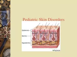
BURNS: cause between 3000 and 4000 deaths/yr in the US and about 2 million physician visits. Burns: thermal, radiation, chemical, or electrical contact. Classified by depth- 1st-degree- affect only the epidermis, causing reddening of the skin, pain, and edema. 2nd -degree- affect not only the epidermis but also some of the dermis, causing reddening of the skin, acute pain, and the formation of blisters and edema. Heals by scarring 3rd -degree- destroy the full thickness of the skin (epidermis, dermis, subcutaneous fat, muscle, and bones. Requires skin grafting and by percentage of total body surface area (BSA) involved – fatal if- Burns of > 40% of BSA Age > 60 yr or < 2 yr Presence of simultaneous major trauma or smoke inhalation
Most of the third finger has a full-thickness burn, where the skin is dark and leathery. The base of the finger has blisters and redness, indicating that this part of the finger has a partial-thickness burn. Partial-thickness (2nd-degree) burn
Thermal burns –Any external heat source (flame, hot liquids, hot solid objects, or, occasionally, steam Radiation burns –Prolonged exposure to solar ultraviolet radiation (sunburn)/ other sources of ultraviolet radiation (eg, tanning beds) or from exposure to sources of x-ray or other non solar radiation. Chemical burns - From strong acids, strong alkalis (eg, lye, cement), phenols, cresols, mustard gas, phosphorus, and certain petroleum products (eg, gasoline, paint thinner). Skin and deeper tissue necrosis caused by these agents may progress over several hours. BURN Categories:
Electrical Injuries Skin burns, damage to internal organs and other soft tissues, cardiac arrhythmias, and respiratory arrest. Higher the voltage (V) and amperage, the greater the ensuing electrical injury Treatment Shut off current Resuscitation Analgesia Sometimes cardiac monitoring for 6 to 12 h Wound care BURN Categories:
Moxa and Cancerhttp://www.cancer.org/Treatment/TreatmentsandSideEffects/ComplementaryandAlternativeMedicine/HerbsVitaminsandMinerals/moxibustion • Direct moxibustion can burn the skin. • Oils from mugwort and wormwood can cause toxic reactions if taken internally, although their toxicity is much lower when applied externally. • Mugwort is on the Commission E (Germany’s regulatory agency for herbs) list of unapproved herbs. This means that it is not recommended for internal use because it has not been proven to be safe or effective, due to the possibility that it may cause miscarriage in pregnant women. • Moxibustion can result in burns and may be dangerous for diabetic patients due to reduced sensation and problems with infection.
Acanthosis Nigricans (AN) Heredity/ Endocrine disorder—most commonly associated with- insulin resistance/ hyperandrogenic state/ hypothyroidism/ obesity Drugs—e.g., nicotinic acid, niacinamide, oral contraceptives, steroids Malignancy: usually adenocarcinoma—e.g., gastrointestinal (60% stomach), lung, breast Physical: Hyperpigmented velvety, typically symmetrical plaques predominantly on the neck, axillae, and groin. Investigations: Screen for diabetes Tx options:Topical tretinoin, ammonium lactate, laser therapy, dermabrasion.
Acne Vulgaris: Acne Vulgaris:
Etiology: generalized or focal- palmar, palmoplantar, axillae and affecting 2%—3% of population; most common in adolescence and young adults. Generalized - infectious (e.g., TB), endocrine, or neurologic; Focal hyperhidrosis- cause unknown. Diagnostic criteria for primary focal idiopathic hyperhidrosis: ■ Focal, visible, excessive sweating of at least 6 mo duration without apparent cause with at least 2 of the following characteristics: ■ Bilateral and relatively symmetrical sweating. ■ Frequency of at least 1 episode per wk. ■ Impairment of daily activities. ■ Age at onset 25 yr. ■ Positive family history. ■ Cessation of sweating during sleep. . Investigations: Starch iodine test can be used to outline the area of excessive sweating. DDx:Thyrotoxicosis, medication-induced hyperhydrosis, pheochromocytoma. Management: rule out— infections, malignancy (ask about night sweats). Topical: Aluminum chloride hexahydrate solution in ethanol (e.g., Drysol®), glycopyrrolate iontophoresis. Systemic: Anticholinergics (e.g., Robinul® 1 mg bid—tid), diltiazem, clonidine, Botulinum toxin (Botox®) injections— very effective; can last 6—12 mo. Surgery: Endoscopic thoracic sympathectomy, subcutaneous liposuction Hyperhidrosis
Etiology: Self-limited viral infection of skin caused by poxvirus affecting mainly children. History: Asymptomatic; occasional perilesional pruritus. Physical: 2- to 6-mm flesh colored, dome-shaped, umbilicated pearly papules; most common affected sites—trunk and face of children, genitals/inner thighs of sexually active adults. Course: Lesions typically involute spontaneously within 9-12 mo; may develop adjacent eczema (in 10%). Investigations: Clinical diagnosis, biopsy ifuncertain. Management ■ Observation. ■ Liquid nitrogen cryotherapy. ■ Curettage; may be uncomfortable. ■ Papule incision with a scalpel blade or at home sharp fingernail and expression of contents. ■ Topical cantharidin: Blistering (intentional) will occur; apply with wooden end of cotton swab and cover with tape for 30 min, then wash off with soap and water; ideal in children. ■ Imiquimod cream. ■ Tretinoin cream. ■ H2RA – Zantac (40 mg/kg/d x 2 mo). ■ Testing for other STDs recommended in adults with lesions in the genital area. Molluscum Contagiosum
Etiology: Infestation by wingless, 6-legged insects Spread by direct contact or through fomites (e.g., clothing, bedding). History: Symptoms none to extreme pruritus. Pediculosis capitis (head louse) Gray-white nits/eggs or lice firmly adhere to hair shaft; postauricular and occipital regions commonly affected; children adults. Pediculosis pubis (crab louse) Eyelashes and pubic hair may be affected; usually transmitted by sexual contact; nits seen on hair, red excoriated skin. Body lice Infest individuals with poor hygiene or those living in crowded conditions; eggs commonly found along seams of clothing. Investigations: Magnifying glass—assisted observation of nits or mature lice. DDx: Impetigo, insect bites, scabies. Management ■ Head lice: Wash hair, rinse, and towel dry; then apply permethrin cream (Nix®, Elimite®) rinse for 10 min and rinse out; repeat in 7 d. ■ Pyrethrin, malathion, crotamiton, petrolatum. ■ For pediculosis pubis—affecting eyelashes, apply petrolatum to eyelashes bid—tid x 10 d. ■ Nits may be removed with vinegar soaks (vinegar:water 1:1) and a fine-toothed comb. ■ Soak combs and brushes in permethrin shampoo for 10 min or boil. ■ Bedding, clothing, and head gear should be washed and heatdried; environment should be vacuumed; unwashable items can be sealed in plastic bags for 2 wk. ■ Contacts should be treated similarly. ■ Ivermectin (oral) increasingly used in resistant or complicated cases of lice & scabies. Pediculosis (Lice)
Common chronic inflammatory disorder of the face. Role of Demodex mite controversial. Easy and recurrent flushing, exacerbated by heat \ (shower, hot drinks), spicy foods, sunlight, cold, alcohol, stress. Sensitive skin. May complain of dry and gritty eyes. Peak incidence 30—50 yr; Physical: Erythema, telangiectases, papules, and pustules of central face; no comedones in contrast to acne. Sebaceous hyperplasia, seborrheic dermatitis & facial lymphedema more common. 4 Major Subtypes Erythematotelangiectatic, papulopustular, ocular, phymatous. Chronic inflammation may progress to rhinophyma (enlarged nose; in males). Ocular involvement common (e.g., gritty, conjunctival injection, styes, photophobia). Investigations: Clinical diagnosis; uncommonly, skin biopsy to rule out lupus or sarcoidosis. DDx: Acne, lupus erythematosus, perioral dermatitis, sarcoidosis, seborrheic dermatitis. Management ■ Based on severity and subtype. ■ Lifestyle modification: Avoid triggers; sun protection & avoidance; facial massage for lymphedema. ■ Topical antibiotics ■ Metronidazole 0.75% gel or 1% cream bid. ■ Sodium sulfacetamide lotion 10% bid. ■ Oral antibiotics (moderate to severe cases with inflammatory papulopustular component): ■ Tetracycline 500 mg po bid ■ Minocycline 100 mg po od—bid. ■ Doxycycline 20 mg po bid (subantimicrobial dose therapy) or 100 mg po qd–bid. ■ Isotretinoin (low dose); less commonly, topical retinoids may be used. ■ Laser therapy (e.g., PDL, IPL) for telangiectases & ablative laser (e.g., CO2) for rhinophyma. ■ Camouflage makeup (e.g., Dermablend™, Covermark®) for erythema. ■ Ophthalmologist to assess for ocular involvement (blepharitis, conjunctivitis, episcleritis). Rosacea
Leg Ulcers • Etiology: Most common lower extremity ulcers are: venous, arterial or neuropathic; can be mixed. Other causes: Trauma, vasculitis, pyoderma gangrenosum, bacterial infections, malignancy, sickle cell anemia). • History: Ask about—onset & course, symptoms, PMHx, social history, & meds. • Physical: Inspect & document—location, size, shape, odor, ulcer edge & base features, surrounding skin (cellulitis, dermatitis), peripheral pulses, & capillary refill. • Investigations: Vascular studies, blood tests, culture & biopsy wound edge; bone scan if you suspect chronic process or osteomyelitis. • DDx: Pyoderma gangrenosum, vasculitis, SCC.
Principles of Wound Care Treat any underlying cause: ■ Venous insufficiency—compression. ■ Arterial insufficiency—surgery. ■ Neuropathic or decubitus ulcer—pressure relief. ■ Infection—antibiotics. ■ Neoplasm—surgery, radiation. ■ Promote wound healing. ■ Débride: Enzymes,surgery/ curette.Dressings: Promote moisture, but not oozing/ weeping; know your dressings— foams, alginates, hydrogels, hydrocolloids, hydrofibers. Infection control: Topical or oral Antibiotics. Skin grafting. Less commonly: skin substitutes, growth factors. Allergic contact dermatitis common with long-term topical antibiotic use (esp. bacitracin, neomycin); use petroleum jelly— low allergenicity, safe, & cheap. Compression stockings required if evidence of pitting/stasis dermatitis; obtain ankle-brachial index prior to initiatingtreatment; medical grade better than over-the-counter. Topical metronidazole eliminates odor. Short-contact topical tretinoin appears to be beneficial. Biopsy ulcer if fails to improve with care. Ulcers, Leg- Management
Acquired skin disorder characterized by well defined areas of complete epidermal depigmentation; various pathogenesis theories; likely a large autoimmune component. History: Asymptomatic; 50% present before age 20; 1%—2% affected; rare in infancy & old age. Associated with immune disorders; therefore work-up for thyroid disease (TSH), diabetes, pernicious anemia, and other autoimmune diseases occasionally indicated (e.g., family history). Course: increased risk of sunburn and skin cancer in amelanotic areas; frustrating to Tx. Investigations: TSH and fasting blood glucose. Vitiligo
DDx: Leprosy, pityriasis alba, tinea versicolor, tuberous sclerosis, Management Mainly for cosmetic/psychological burden; consider assessment for Diabetes, Thyroid & Addison disease. Topical steroids and new topical immuno modulator (tacrolimus & pimecrolimus), PUVA, NB-UVB phototherapy, camouflage makeup; oral steroid rarely used. Note that facial and more proximal lesions respond better than acral lesions. ■ Surgical transplants can be considered in stable vittiligo— uncommon ■ If extensive, rarely can consider bleaching of non affected areas to result in total white color. ■ Advise re: importance of sun protection. ■ Autologous transplantation or grafting of cultured autologous melanocytes in stable vittiligo (still in research) Management of Vittiligo
Beau’s Lines • Deep grooved lines that run from side to side • Look like indentations • Temporary cessation of cell division in the nail matrix • Infections, coronary artery disease, hypocalcemia • Beta blockers • Malnourishment
Muehrcke lines • Paired, white, transverse lines that signify an abnormality in the vascular bed of the nail seen in √chronic hypoalbuminemia √ chemotherapy-induced nail changes √ pellagra √ Hodgkin disease √ renal failure √ sickle cell anemia √ nail damage from paraquat
Mee’s Lines • Poisoning with arsenic, thallium or other heavy metals, and can also appear if the subject is suffering from renal failure
‘Half and Half’ nails • Typical in chronic renal failure and uremia
Leukonychia (White nails) √ hypoalbuminaemia √ nephrotic syndrome √ liver failure, √ protein malabsorption ad √ protein-losing enteropathies
Nail beds in SLE/ Dermatomyositis √ Nail bed vasculitis √ Copper colored rash
Onycholysis-spontaneous separation of the nail √ seen in women with long fingernails √ common cause: trauma
Splinter hemorrhages √ Trauma to the nail √ Subacute or acute bacterial endocarditis