Week 6: Cell Morphology
Wright stain RBC morphology Anisocytosis Poikilocytosis WBC morphology. WBC differentials Artifacts. Week 6: Cell Morphology. Wright Stain. Romanowsky stain family Polychromatic Absolute methanol fixative Methylene blue - basophilic components Eosin - acidophilic components.

Week 6: Cell Morphology
E N D
Presentation Transcript
Wright stain RBC morphology Anisocytosis Poikilocytosis WBC morphology WBC differentials Artifacts Week 6: Cell Morphology
Wright Stain • Romanowsky stain family • Polychromatic • Absolute methanol fixative • Methylene blue - basophilic components • Eosin - acidophilic components
Smear Making • Essential in morphology • Wedge method • Thickness and length
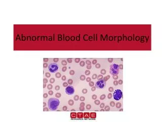
Key Features • Size • Nuclear shape • Chromatin features • Granules and other inclusions • Color
Leukocytes • Granulocytes: have specific granules • Eosinophil • Basophil • Neutrophil • Non-granulocytes • Lymphocyte • Monocyte
Granulocytes • Eosinophil • Large orange granules • <2% in circulation • Usually bilobed • Increased in allergy, parasites
Granulocytes • Basophil • Large black-purple granules • <1% in circulation • Increased in allergy, CML
Granulocytes • Segmented Neutrophil • Small purple granules • 54-62% in circulation • Coarse chromatin • Increased in bacterial infection • Band • No nuclear filament • 3-5% in circulation • Increased in bacterial infection
Non-Granulocytes • Lymphocyte • Relatively small • Round or oval nucleus • Smudged chromatin • Basophilic cytoplasm • Increased in viral infection • 25 - 33% (more in children)
Non-Granulocytes • Monocyte • Relatively large • Grey cytoplasm with vacuoles • Irregular nucleus • Linear chromatin • Increased in chronic infection • 3 - 7%
Platelets • Cytoplasmic fragments of large megakaryocyte • 2-3 mm in diameter with granules • Promotes clot formation • 8-20 per oil immersion field • 150,000 - 400,000/mL
Erythrocyte Morphology • Normal: biconcave disc of 7.5 mm diameter • Has central pallor (1/3 of diameter) on smear • No inclusions
Anisocytosis: Variation in Size Microcytes Macrocytes
Poikilocytosis: Abnormal Shape Target Cells Hypochromic
Poikilocytosis Schistocyte Sickle Cell
Poikilocytosis Teardrop Cell Spherocyte
Poikilocytosis Elliptocyte Stomatocyte
RBC Inclusions, etc. Polychromasia Howell-Jolly Bodies
RBC Inclusions Cabot Ring Basophilic Stippling
RBC Inclusions, etc. Malaria Rouleaux
Too Thick Too Alkaline Too Thin Too Acidic