Download

1 / 21
230 likes | 483 Vues
More on smFRET and high-resolution microscopy. You give a (brief) talk? (4 per class: 10 minute.). Apr 21 Apr 28 May 5. Last time: smFRET. Have problems with getting 100% labeling Have problems synchronizing. G-quadruplex: at least 3G, repeated 4 times.

E N D
You give a (brief) talk?(4 per class: 10 minute.) Apr 21 Apr 28 May 5
Last time: smFRET Have problems with getting 100% labeling Have problems synchronizing. G-quadruplex: at least 3G, repeated 4 times
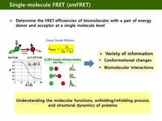
Another reason to do smFRETLabeling is stochasticImagine protein is made up of 5 identical subunits, want 1D, 1A Require: ONEDonor ONEAcceptor onONEChannel D: Alexa 488 Reality: Labelingisstochastic. (evenwith optimization) A: Alexa 568 Mechanosensitive Channel
Virtually Sort Out Channels with 1D & 1A by Photobleaching Use photobleaching to count the number of attached fluorophores. Rf Donor trace Donor trace How many distances expect to see? E? Wang et al. eLife, 2014 Rn
Labeling Stoichiometry 2 donor’s 1 donor, 1 acceptor There is direct excitation; subtracted out. Note: Donor : Donor & Acceptor-Acceptor, thrown away D:nA ; nD:A ; nD:nA ; also gotten rid of by photobleaching
Eukaryotic MSCs Hearing Garcia et al, J. Neurosci, 1998 Corey et al, Nature, 2004 Cell growth (tumor metastasis) Lichtman et.al, Hematology Zarychanski et al, Blood, 2012 Coste et al, Nature, 2012 Touch Kirber et al, Pflugers Arch, 1988 Simon et al, J. Physio, 2009
Mechanosensitive Channels of Large Conductance (MscL) • Last “safety valves” • Large nonselective pore • High conductivity ~ 3 nS • Crystalized in the closed state (Mycobacterium tuberculosis) • Homo-pentamer • Each subunit: TM1, TM2 and CP • TM1 & TM2 for gating; CP as a pre-filter Chang et al, Science, 1998 BUT: NO crystal structure for the open state!
Distribution of FRET efficiency (E) Expect 2 E, for closed; 2 E’s for open Why are there THREE peaks? M42C some open some closed open Closed (~23% open) closed Open E = 0.23 Primarly opened Closed (not all opened) After adding LPC, the high-E peak decreases, clearly showing the opening of the channels. Closed (not all opened) Wang et al. eLife, 2014
How does MscL open? Helix-Tilt Model Barrel-Stave Model Membrane TM1 Perozo et al, Nat Rev Mol Cell Biol, 2006 TM2
Other techniques for high-resolution (besides STORM/PALM) STED, 2-Photon microscopy
STochastic Optical Reconstruction Microscopy PhotoActivation Localization Microscopy STORM & PALMMost Super-Resolution MicroscopyInherently a single-molecule technique Zhuang, 2007 Science Betzig, 2006 Science 1. Activation (405 nm) (widefield) 2. Fluorescence ~ 500-650nm (widefield) Huang, Annu. Rev. Biochem, 2009 Cy3-Alexa 647 2-color secondary antibodies Cy2-Alexa 647
1 mm 1 mm 1 mm Correlative PALM-EM imaging PhotoActivation Localization Microscopy (F)PALM (Photoactivatable GFP) TIRF PALM EM Mitochondrial targeting sequence tagged with mEOS Patterson et al., Science 2002
center width STORM/PALM: Get fantastic resolution (1-30 nanometers) by repeatedly localizing point-spread-function very accurately (based on FIONA) Collect from ~ 1-10k photons STimulated Emission Depletion: STED Get fantastic resolution (6-50 nanometers) by narrowing point-spread-function. Downside:
STimulatedEmission Depletion (STED) Recent development in super-resolution microscopy S. Hell Net result is a smaller Point Spread Function Sharpen the fluorescence focal spot is to selectively inhibit the fluorescence at its outer part. Make the molecules lase by using high-power STED beam! 200nm http://www.mpibpc.gwdg.de/groups/hell/ Huang, Annu. Rev. Biochem, 2009
Biological Example of STED The transient receptor potential channel M5 Analysis of spot size for Confocal (A) and STED (B) images of TRPM5 immunofluorescence layer of the olfactory epithelium. (A, C Inset) Confocal image at a lower (higher; box) magnification taken with a confocal microscope. (B) STED image. Effective point-spread function in the confocal (189 nm) and STED (35 nm) imaging modes. Hell, PNAS, 2007
Two-Photon Microscopy (Watt Webb, Science, 2003) You get Z-axis for free!Automatic confocal detection with 2-photon microscopy…plus other advantages
Two-Photon Microscopy Inherently confocal, long wavelength (less scattering) two-photon One-photon emission Intensity 2p 1p wavelength Simultaneous absorption of two photons Reasonable power if use pulsed laser
One photon two photon (Dis-)Advantages of 2-Photon Excitation objective Inherent spatial (z-) resolution Low light scattering (scattering like l-4) Single-color excitation with multiple emission colors Disadvantage: Huge Instantaneous Excitation Powers: must use photostable dyes (e.g. quantum dots)
2-Photon Widefield Excitation of Single Quantum Dot • Blinking and emission intensity – laser power plot prove that it is single Qdots and 2-photon excitation Qdot585, 655 in PBS buffer, no reductants (no deoxygenation) <P> = ~150 W/cm2 , 30 msec/frame, scale bar 1 um. 160 nm effective pixel size 50x lower power withSingle Quantum Dot than with single fluorophores 46 mW 449 mW