Download
1 / 36
360 likes | 391 Vues
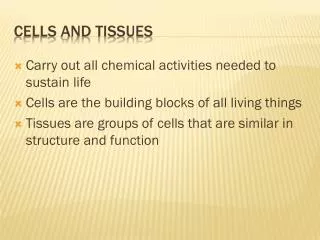
Compartmentation: Cells and Tissues. 3. Three Major Body Cavities. The body is divided into various cavities but not all compartments have walls or are completely enclosed. Figure 3-1. Lumens of Hollow Organs.

E N D
Three Major Body Cavities The body is divided into various cavities but not all compartments have walls or are completely enclosed Figure 3-1
Lumens of Hollow Organs • Hollow organs- contain a space filled with something other than the organ’s tissue. • Heart • Lungs • Blood vessels • Intestines • Lumen – interior of a hollow organ • Fluid-filled interior • Not the internal environment- as is in the GI tract
Functional Compartments • Extracellular fluid- found outside of organ tissue • Plasma-fluid of blood • Interstitial fluid- fluid between blood vessels and tissue cells • Intracellular fluid-fluid inside tissue cells
Body Fluid Compartments Figure 3-2
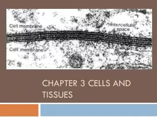
Cell Membrane: Overview Membranes in the body may be macroscopic or microscopic and serve different functions Figure 3-3
Cell Membrane: Function • Physical barrier- separates intracellular and extracellular fluid • Gateway for exchange- controls what enters and leaves the cell • Communication- surface proteins respond and recognize other molecules which can change cell activity • Cell structure- cell shape is maintained by cytoskeletal proteins attached to membrane proteins. Membrane proteins also form cell junctions • Phospholipid bilayer- composed of mostly lipids and proteins, it’s hydrophobic and hydrophilic regions assist in controlling transport.
Cell Membrane: Structure The fluid mosaic model of a biological membrane Figure 3-4
Cell Membrane: Composition Lipids Phospolipids – a glycerol molecule with one phosphate and two fatty acid tails- makes up a large percentage of the membrane. Cholesterols- imbedded in the bilayer it stabilizes the membrane and reduces it’s freezing point. Proteins Integral – transmembrane protein, serves as a channel Peripheral – side proteins that may be enzyme of cytoskeleton anchors Lipid-anchored – associate with sphingolipids to form lipid rafts that may attract other proteins or enzymes Carbohydrates Glycolipids- carbohydrates and fatty acids Glycoprotiens-carbohydrates and proteins
Cell Membrane: Formation Phospholipid molecules are composed of two fatty acid chains, one glycerol molecule, & one phosphate group Figure 3-5a
Cell Membrane: Formation Figure 3-5b
Cell Membrane: Proteins The three types of membrane proteins: integral, peripheral, and lipid-anchored Figure 3-6
Cell Membrane Concept Map of cell membrane components Figure 3-9
The cell membrane covers cells of various sizes, shapes, and functions Figure 3-10
Cell Compartments • Cytoplasm- The space between the plasma membrane and the nucleus • Cytosol - the jelly like substance that suspends the organelles • Inclusions - a non-membranous organelle or insoluble particles • Organelles - cell structures with specific function- “small organs” • Nucleus- contains the genetic information for the cell as chromatin, the nucleolus, and nucleoplasm.
Cell Compartments A map for the study of cell structure Figure 3-11
Organelle “Factory” and summary chart • See board drawing and table on board • Review on your own the functions and structures of the following cell organelles (see fig 3:12): • Inclusions (3-types) • Centrioles, Cillia, & Flagella • Cytoplasmic protein fibers (3 sizes) • Cytoskeleton • Mitochondria • Smooth/Rough Endoplasmic Reticulum • Cytoplasmic Vesicles • Nucleus
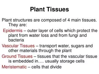
Primary Tissue Types • Epithelial- major functions: are protection, exchange, and lining cavities. • Connective- major functions are: support, storage, communication, immunity. • Muscle- major functions are: posture, movement, heat production, support and protection. • Nerve- major function is communication and control, information processing.
Epithelial Tissue: Structure • Basal lamina • Basement membrane
Epithelial Tissue: Function • Exchange – quick movement of molecules • Transport – move from one side to another and process • Ciliated – move substances in the extracellular matrix • Protective – multiple layers, quickly regenerates • Secretory – produces substances secreted into the extracellular matrix or outside the body.
Exchange Epithelia • Single cell layer of flat cells that allow molecules to cross through at different rates, increase surface area with microvilli. • Rapid transport - • Oxygen - • Carbon dioxide - • Ions and fluids - • Capillaries and lung alveoli -
Transporting Epithelia • single layer or cuboidal or columnar cells, take in a molecule from a lumen and transport it into the blood stream. • Exchange of ions and nutrients - • Tight junctions - • Intestine and kidney -
Ciliated and Protective Epithelia • apical cilia allow more the movement of substance on the surface of the cell, like the ovum or mucus • Ciliated epithelium - • Trachea - • Sweep mucous out - • Protective epithelium - • multiple layers and in skin, serve for protection. Cell have a high regenerative ability. • Skin - • Prevent exchange -
Secretory Epithelia • contain goblet cells and cells that form the different endocrine or exocrine glands in the body • Exocrine tissues • Mucous glands • Goblet cells • Secreted externally via ducts • Endocrine tissues • Hormones • Secreted to ECF and blood
Secretory Epithelia Development of endocrine and exocrine glands from epithelium Figure 3-28 (1 of 3)
Connective Tissues: Structure • Support and barriers – strong high collagen content allow to withstand forces • Ground substance – varies in amount of water and changes the consistency of the type of connecitve tissue • Cells – have a wide variety of functions • Fixed – imbedded in a dense ground substance • Mobile – blood cells surrounded by a fluid ground substance such as plasma, are able to enter or leave the blood stream.
Connective Tissues: Structure • Fibers and their functions- found in the ground substance, the different ratios of each give each type of connective tissue their unique characteristics. • Fibroblast cells - produce the fibers and ground substance • Collagen – has a stronger tensile strength than steel, there are 12 variations, is most abundant in the body. • Elastin – gives elasticity to tissues • Fibrillin – combines with elastin to give support to elastic organs. • Fibronectin – stick to extracellular matrix of cells and helps in forming blood clots • Reticular fibers- form a network of supportive fibers for cells composed of free cells as in bone marrow, spleen, and lymphnodes
Cells and Fibers of Loose Connective Tissue Figure 3-29 (1 of 2)
Various Connective Tissue Types • Strength or flexibility • Tendons and ligaments • Collagen dominates • Adipose connective tissue • White • Single droplet • Brown • Multiple droplets • Blood • Plasma matrix • Free blood cells • Cartilage • Light and flexible • Trachea and ears • Bone • Calcified • Rigid
Muscle Tissues • Contractile • Force and movement • Signal conduction • Types • Cardiac • Smooth • Skeletal
Nervous Tissues • Neurons send signals • Excitable • Electrical • Chemical • Glial cells support
Cell Death and Replacement • Apoptosis- cell death not caused by injury or other external reasons • Normal cell replacement – during body formation, or in normal body function cells reach a life limit and die • Programmed cell death - induced by the cell without disturbing adjacent cells; “cell suicide” • Stem cells – undifferentiated cells that can become any cell needed in the body, totipotent, puripotent, and mulitpotent • Role in cell replacement – certain tissues have multipotent stem cells that can replace cells • Research uses and potential – need to find a good source of stem cells, face many ethical issues
Organs • Groups of tissues with related function – each contains the four types of tissues in various ratios • Epidermal tissue (skin) - • Multiple cell layers – epidermis, dermis, hypodermis • Multiple tissue types – epitheial, connective, muscular, nervous • Multiple functions – protection, metabolism, temperature regulation, water proofing, blood storage, insulation, excretion, sensory organ
Integument System Functions • Protection • Insulation • Water proofing • Temperature regulation • Excretion • Cutaneous Sensory organ • Metabolism • Blood reservoir