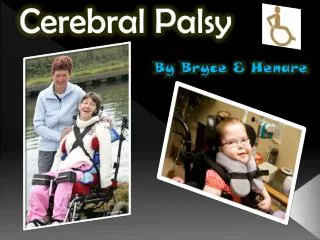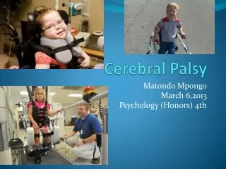
Orthopaedic Considerations in Cerebral Palsy
Orthopaedic Considerations in Cerebral Palsy. Stewart Morrison Western Health Friday Presentation 20 th January 2012. Definition + Aetiology. “ a disorder of movement and posture due to a defect or lesion in the developing brain ”

Orthopaedic Considerations in Cerebral Palsy
E N D
Presentation Transcript
Orthopaedic Considerations in Cerebral Palsy Stewart Morrison Western Health Friday Presentation 20th January 2012
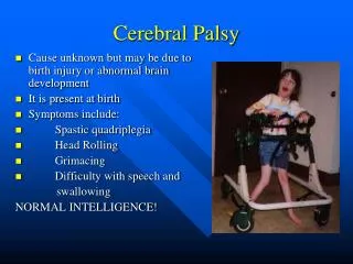
Definition + Aetiology “a disorder of movement and posture due to a defect or lesion in the developing brain” Not a diagnosis, but a heterogenous collection of clinical syndromes Cerebral lesion is static, musculoskeletal pathology is progressive Prenatal placenta insufficiency, toxins, genetic factors, TORCH Perinatalpremature delivery, hypoxia, infection, kernicterus, haemolytic disease Postnatal infection, trauma
Classification Type of Motor Disorder Spastic pyramidal system (motor cortex) Athetoid extrapyramidal (basal ganglia) Ataxis cerebellum + brainstem Rigid basal ganglia + motor cortex + Mixed Limbs Involved Monoplegia one limb (rare) Hemiplegia one side Diplegia lower limbs, assymetrically Triplegia three limbs (rare) Quadriplegia four limbs
Demographics Two per 1000 live births 50% have normal intelligence, 25% able to self-support as adult Incidence remains static +/- increasing
Clinical Features I Dependent on: • Severity of neurological lesion • Location of neurological lesion • Age of child • Absence of normal reflexes (blinking, sucking) • Persistence of abnormal reflexes (Moro’s reflex) • Delayed motor milestones (head control 3 months, sitting 6 months, walking 12 months) • Gait disturbance • Epilepsy, speech and hearing difficulties, visual defects, feeding difficulties, drooling, learning, behavioural problems
Clinical Features II Posturing sitting (hypotonic slump) standing (crouchposture, spastic posture, pelvic obliquity, loss of lumb. Lordosis) Gait athetoid or ataxic movement Neuromuscular UMN or spastic paresis resistance to passive movement Babinski +ve Deformities Equinus FFD Knee
Pathology I • Skeletal muscle growth depends on regular stretching of relaxed muscle, under physiological loading • In CP: • Muscle does not relax (spasticity) • Reduced activity (weakness + balance)
Pathology II • Dynamic Contractures correctable deformity • Muscle Contractures fixed deformity • Secondary Bone Changes e.g. medial femoral torsion, lateral tibial torsion
Management Concepts Limitations • Treating the sequelae of a neurological lesion, not the lesion itself • Many of the operations were developed for the management of polio myelitis Stage I Physiotherapy, Orthotics, Botulinum Toxin, Selective Posterior Rhizotomy Stage II Timing critical and controversial Unpredictable results Staged vs. single procedures Stage III Correctional osteotomies for torsional + joint deformities
Tendon Transfer: Principles • Correct joint contractures • muscle of adequate strength • muscle of adequate excursion • one tendon for one function • an expendable donor • a straight line of pull • Position and time transfers so that they lie in tissue of optimal condition
Lower Extremity I Age of surgery critical • Gait evolves into adult pattern by age seven years • Gait deterioration during adolescence is quite common Preoperative evaluation • Multiple joint evaluation required • Eg. TA correction in presence of tight hamstrings will result in persistent crouch at knee and calcaneus gait • Gait Analysis critical • Swing-phase foot clearance, foot progression angle
Lower Extremity II Hemiplegia Group I mild foot-drop gait leaf-spring AFO Group II equinus gait stretching casts, botulinum toxin, AFO, lengthening Group III Knee, medial hamstrings, gastroc recession, medial hamstring lengthening, quadricepts involvement distal rectus femoris transfer Group IV Hip flexion, medial torsion lengthening psoas, external rotation osteotomy, and above Spastic Diplegia Most achieve good function Hip flexors, adductors, medial rotators, calf most affected Secondary bone torsional problems
Lower Extremity III Lengthening Achilles Tendon overused “a little equinus is better than calcaneus” ? Silveskiod Test (Gastroc vs. Soleus) Gastrocnemius Recession Z Lengthening or Percutaneous Techniques Varus Deformity of the Foot Tib Post usually resonsible (stance and swing) Tib Ant (swing only) Lengthening vs. transfer Valgus Deformity Lengthening, Fusion, Osteotomies
Lower Extremity IV Knee Flexion Contracture “crouch” Surgical lengthening of medial hamstrings consideration of NV bundle in severe contracture Stiff-Knee Gait may occur if rectus femoris co-spasticity Rectus Femoris transfer indicated Hip Flexion Contractures often secondary to knee/ankle issues Thomas or Staheli tests Psoas lengthening
Lower Extremity V Hip Subluxation Rotational Osteotomies Hip Reconstructive Surgery (spastic quadriplegia)
Upper Extremity Evaluation • Sensation • Electromyography Principles • Define goals • Restore • Rebalance
Upper Extremity Shoulder • Internal rotation, adduction common Botulinium type A Supscapularis, Pec Major lengthening External rotational osteotomy Elbow • Static and dynamic flexion contractures flexor release dependent on NV bundle Wrist/Digits • Wrist flexion +/- pronation, ulnar deviation lengthening and transfer procedures
Thank you BARCZYNSKI, A., PASIERBEK, M., GAZDZIK, T. S. & KLOSA, Z. 2002. Management of foot deformity in cerebral palsy.Ortop Traumatol Rehabil, 4, 21-6. GRAHAM, H. K. 2005. Classifying cerebral palsy. J Pediatr Orthop, 25, 127-8. KAROL, L. A. 2004. Surgical management of the lower extremity in ambulatory children with cerebral palsy. J Am Acad Orthop Surg, 12, 196-203. GRAHAM, H. K. 2003. Musculoskeletal Aspects of Cerebral Palsy. Journ. Bone & Joint Surgery (British). 85-B, 2:157 SAEED, W. R. 2003. Cerebral Palsy of the Upper Extremity: A Surgical Perspective. Current Orthopaedics.17:105-116