Download
1 / 44
480 likes | 942 Vues
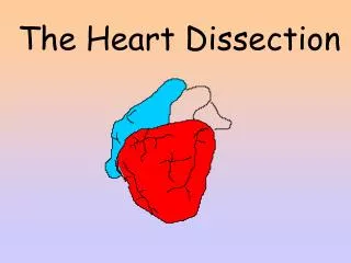
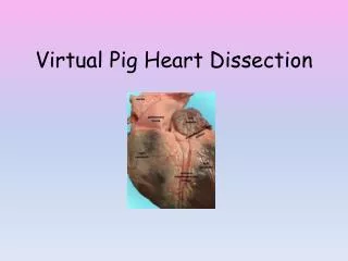
HEART DISSECTION LAB. Procedure. Obtain a dissection pan and dissecting kit. Procedure. Obtain a heart and rinse off the excess preservative with tap water. Pat the heart dry. Place the heart in a dissection pan.

E N D
Procedure • Obtain a dissection pan and dissecting kit.
Procedure • Obtain a heart and rinse off the excess preservative with tap water. Pat the heart dry.
Observe the pericardium. If the pericardial sac is intact then remove the outer layer from its attachment points.
Carefully pull the visceral pericardium (epicardium) away from the myocardium
Examine the external surface of the heart. Notice the accumulation of adipose tissue. This adipose usually accumulates along the boundaries of the heart chambers and along the coronary arteries. Remove as much adipose as possible.
Now you should be able to identify the apex (bottom left "point" of the heart) and the auricles (earlike flaps projecting from the atria).
Locate the pulmonary trunk and the aorta on the superior aspect of the heart. Clear the adipose away from these arteries. The pulmonary trunk divides into the left and right pulmonary arteries. The aorta will have a large branch coming from beneath the pulmonary trunk.
Make a coronal (frontal) cut through the heart. Stop cutting when your knife reaches the top portions of the atria.
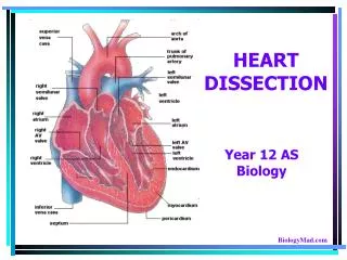
Notice that the heart is made up of three histological layers: the epicardium, the myocardium and the endocardium
Locate the side with the thickest myocardial wall. This will orient you to the left side of the heart
You should see that there are spaces (or "chambers") on the left and right sides of the lower heart.These are the left and right ventricles
You should also see a thick structure dividing the two ventricles, the bulk of which is comprised of cardiac muscle. This is the interventricular septum.
The ventricles are divided from the chambers directly above them by atrioventricular (or "AV") valves. These valves have flaps (or "cusps") to which "heart strings" attach.
The left AV valve has two cusps, so it can be referred to as being a "bicuspid" valve. The right valve has three cusps, so it can be referred to as being a "tricuspid" valve.
The strings that attach to the AV cusps are called chordae tendinea.
The chordae tendineae are anchored to the ventricular walls via papillary ("nipple-like") muscles.
Remove the right ventricular wall and cut into the pulmonary trunk in order to view the pulmonary semilunar valve.
Remove the left ventricular wall and cut into the aortic trunk in order to view the aortic semilunar valve.
1) Adipose Tissue 2) Myocardium of the Interventricular Septum 3) Myocardium of the Left Ventricular Wall
4) Right Ventricle (the space below the right A.V. valve) 5) Left Atrium (the space above the left A.V. valve) 6) Left Ventricle (the space below the left A.V. valve)
7) Aortic Valve (or left semilunar valve) 8) Left Atrioventricular (AV) Valve (which has two flaps, or is "bicuspid"; sometimes also called the mitral valve) 9) Apex (point) of the Heart
10) Ascending Aorta (the tube above the aortic valve) 11) Left Atrium (the space above the left A.V. valve) 12) Chordae Tendineae (the "heart strings" that run from the cusps of the A.V. valves to the papillary muscles)
15. 14. 16.
17. 18. 19.
20. 21. 22
23. 24. 25.