
Physiological & Biochemical Process regulating Parturition
Physiological & Biochemical Process regulating Parturition. Physiological process in human pregnancy that result in initiation of parturition & onset of labor : poorly defined Retreat from pregnancy maintenance & uterotonin induction of parturition hypotheses

Physiological & Biochemical Process regulating Parturition
E N D
Presentation Transcript
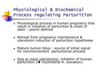
Physiological & Biochemical Process regulating Parturition • Physiological process in human pregnancy that result in initiation of parturition & onset of labor : poorly defined • Retreat from pregnancy maintenance & uterotonin induction of parturition hypotheses • Mature human fetus : source of initial signal for commencement parturitional process • One or more uterotonins: initiation of human parturition ( myometrial R. elevation)
Anatomical & physiological consideration of myometrium • Characteristics – advantage in efficiency of uterine contractions & delivery of fetus ① degree of shortening of smooth m cells with contraction : magnitude greater than in striated m cells ② forces can be exerted in smooth m cells in any direction ③ not organized in same manner as skeletal m - thick & thin filaments in long, random bundles → greater shortening & force-generating capacity ④ multidirectional force generation
Regulation of myometrial contraction & Relaxation • Interaction of myosin & actin activation of adenosine triphosphatase ATP hydrolysis force generation (by enzymatic phosphorylation of MLC) • Ca bind to calmodulin activate myosin light chain kinase increase in intracellular Ca2+ (transient) • Contraction prolonged inhibition of myosin phosphatase activity by Rho kinase • Uterine activity regulation of contraction –associated protein (CAP) : include channels associaeted with smooth m excitation & contraction , gap junction component, uterotonic stimulatory or inhibitory R
1] Myometrial Gap junctions • Cellular signals transferred between cells through intercellular junctional channels • Communication is extabilished between myometrial cells by gap junctions that facilitate passage of electrical or ionic coupling current as well as metabolite coupling • Consist of two protein ”hemi-channels “ termed connexons hexameric assemblage of type of protein called connexin • Conduit for exchange of small molecule • Gap junctions optimal No : electrical synchrony in myometrium , coordination of contraction ( greater force during labor)
2] Cell surface R as regulator of myometrium • Estrogen & progesteron R, variety of cell surface R that can directly regulate contractile state of cell • Most of heptahelical R in myometrium • Activation of adenylycyclase • G-protein-medicated activation of phospholipase C [Ca2+]) ↑& myometial cell contraction ↑ • In high concentration: from maternal blood (endocrine), contiguous tissues or adjacent cells (paracrine) or direct synthesis in myometrial smooth m cell (autocrine) [Fig 6-16]
2] Cell surface R as regulator of myometrium
2] Cell surface R as regulator of myometrium • Myometrial response to H. can change during course of pregnancy • Imposition of quiescence (activation of adenylyl cyclase) or facilitation of contraction ( activation of phospholipase C & increased [Ca+])
A Fail-Safe system that maintains Ut quiescence • Multiple process act independently & cooperatively to estabilish ut quiescence • To sustain Ut quiescence of phase 0 : biomolecular systems ( neural, endocrine, paracrine, and autocrine )
A Fail-Safe system that maintains Ut quiescence • Phase 0 of parturition & its quiescent state factor • Actions of estrogen & progesterone via intracellular R • Myometrial cell plasma membrane R –mediated increase in cAMP • Generation of cGMP • Other systems, including modifications in myometrial cell ion channels
Several independent pathways, defects in one component of this system not preclude successful maintenance of preg to term [Figure 6-17] key factors to regulate phase of parturition
Progesterone & Estrogen contributions to Phase 0 of parturition • Maintains Phase 0 of human parturition • Removal of Progesterone ( Progesterone withdrawal) : progression of phase 0 into phase 1 of parturition • Progesteron action: • Successful maintenance of preg • Biomolecular evidence or role of other agents not defined • Maintain Phase 0 of human parturition • Estrogen action : • Promote progesteron responsiveness Ut quiescence • In responsive tissues, Estrogen R. induces Progesteron R synthesis
Steroid H Regulation of myometrial Cell-to-Cell communication • Progesteron • Decrease expression of contraction associated proteins • CAP grouping : smooth m excitation contraction, gap junction components uterotonic stimulatory R • Inhibit expression of gap junctional protein connexin 43 • Progesterone antagonist: premature development of gap junction preterm labor & delivery • Connexin 43 mRNA in human myometrial tissue increase before labor between 37 ~40 wks. • Gap junction in myometrium increase • But expression of connexin 43 protein not increase during gestation or at labor ? in intracellular regulator of actual gap junction assembly at time of labor
Heptahelical R that promote myometrial relaxation • Multiple process act independently & cooperatively to estabilish ut quiescence • Associated with Gas-mediated activation of adenyly cyclase & increased level of cAMP in myometrium • Part of fail-safe system to maintain Ut quiescence of phase 0 of parturition
B-adrenoreceptors • B-adrenoreceptors mediate Gas- stimulated increase in adenylyl cyclase increased level of cAMP myometrial cell relaxation • Exact role of catecholamines in maintaining ut quiescence : ill defined
Luteinizing H (LH) & chorionic gonadotropin(hCG) • LH & hCG R in myometrium during preg greater before than during labor • Chorionic gonadotropin(hCG) activate adenylyl cyclase by plasma memb R Gas-linked system • decrease in contraction frequency & force & tissue-specific myometrial cell gap junctions
Relaxin • Peptide H member of insulin like growth factor family of proteins, A & B chain • Secretion from corpus luteum • Greates & peak at 1ng/ml 8wks ~12wks. • Thereafter decline to lower lever until term • Activation of adenylyl cyclase & promotes myometrial realxation, effect cervical softening
Corticotropin–Releasing H(CRH) • Myltiple isoforms their affinity & coupling modified late in preg • Sythesized in PL,amnion,decidua,myometrium • Increase final 6~8wks of preg • Signal through cAMP or Calcium • Relaxation or contraction of myometrial cell depending on R isoform present • CRH role of uterorelaxant during phase 0 & uterotonin in phases 1 & 2 of parturition
Parathyroid H–related protein (PTH-rP) • Initiate Gas-medated activation of adenylyl cyclase • Expressd in myometrium,amnion,decidua & trophoblast • PTH-rP expression in smooth m icreased by m stretch • Function : not established,serve to maximize ut blood flow durng myometrial contraction by vasorelaxant action • Facilitate maintenance of Ut tranquility
Prostaglandins • Interact with family of 8 different heptahelical R • PG : uterotonins,prostanoid sometimes can act as smooth m relaxant • Individual prostanoid : diverse effect
Prostaglandins • By action of phospholipase A2 or C • Arachidonic acid act as substrate of type 1 & type 2 PG synthase (PGHS-1 & -2) called COX –1 & –2 • Both convert Arachidonic acid to unstable endoperoxide PG G2 and then to PGH2target of many NSAIDs & act as tocolytics to prevent preterm labor • PGH2 convert to active PG (PGE2,PGF2 & PGI2) • PGDH Expression :regulate in Ut rapidly incactivate PG metabolites
Prostaglandins • PG family of R classifed according to specificity of binding of given R to particular PG • DP(PGD2) & IP (prostacyclin or PGI2) : increase intracellular cAMP • FP R (PGF2a) : increase intracellular Ca • EP2 & EP4 (PGE2): activate cAMP production • PGE2 PGI2 : maintain Ut quiescence by increasing cAMP signaling • PGE2,PGD2,PGI2 : relaxation of vascular smooth m & vasodilation
Prostaglandins • Either generation of specific PG or relative expression of various PG R determine responses of human myometrium • Change with gestation (32~35 wks vs 39~40wks .) • Regional change in upper & lower ut segment • Prostanoid : myometrial relaxation at one stage of preg & regional myometrial contraction after initiation of parturition (in fundus)
Atrial & Brain natriuretic peptides & cyclic guanosine monophosphate(cGMP) • Guanylyl cyclase activation increase intracellular level of cGMP promote smooth m relaxation • ANP & BNP stimulate intracellular level of cGMP↑ • BNP secreated by amnion,ANP expressed in PL • Soluble form Guanylyl cyclase activated by nitric oxide penetrate pl membrane to enter cell • NO react with iron in Guanylyl cyclase enzyme stimulate to produce cGMP act myometrial relaxation
Accelerated Uterotonin degradation & Phase 0 of parturition • To stimulate myometrial cell refractoriness, Activity of enzyme↑: degrade or inactivate endogenoulsy produced uterotonins • Uterotonins (degredative enz) PG(PGDH), endothelin (enkephalinase), oxytocin (oxytocinase), histamine (diamine oxidase), catecholamines(catechol O-methlytransferase), angiotensin-II (angiotensinase), PAF(PAF –acetylhydrolase) • These enzyme increase by Progesteron action & decrease late in gestation
Fail-safe system for Ut activation • Phase 1 of parturition: morphological & functional change in myometrium & Cx that prepare Ut for labor • Development of uterotonin sensitivity, improved intercellular communicability via gap junctions • Alteration incapacity of myometrial cell to regulate concentration of cytoplasmic Ca2+ • The process leading to enhance uterine responsiveness activation (by Chalis & associates (2000)) • As fuctional contractile capacity of myometrium & Cx ripened, phase1 merge into phase 2 • Alteration in timing of these process cause preterm & delayed labor
Classical Progesteron withdrawal not cause human parturition • In many species, plasma progesterone level decrease • Activation of Ut in preparation for labor • Associated with increase in estrogen level in several species • In primate, plasma progesteron level not decrease before labor, only after delivery of PL decline • Nonetheless, morphological & fuctional modification that prepare Ut for labor occur in timely manner in human
Classical Progesteron withdrawal not cause human parturition • In species, Progesteron withdrawal can be blocked by administering Progesteron to mother • Conflicting reports whether or not Progesteron delay timely onset of parturition or prevent preterm labor • Majority of studies Progesteron cannot prevent preterm labor not appear to extend labor in control group • Progesteron metabolite, 17-hydroxy progesteron (less potent than Progesteron ):minimally decreased incidence of preterm labor in high –risk group • additional research need
Progesteron R Antagonist & human parturition • RU 486, mifepristone: less effective in inducing abortion or labor in later preg , effective in ripening Cx & increase myometrium sensitivity to uterotonins (Chwalisz & Garfield,1994) • Decreased circulating progesterone by inhibitioning enzyme 3B-hydroxysteroid degydrogenase induced labor • Inhibition of progesterone action: important for activation phase of parturition • But there is : ‘hidden’ or unique form of fuctional progesteron withdrawal that end ut quiescence
Fuctional Progesteron withdrawal in human parturition • Unique mechanisms to inhibit progesterone action in human • Late gestational decrease in activity of progesteron R expression that cause fuctional withdrawal • Changes in relative expression of progesterone R or of its two isoforms • shift in relative ratio of PR-A to PR-B(active isoform) within myometrium • Activity of Progesteron R for gene transcription in late gestation • coactivator ↓ • co-repressor ↑
Oxytocin R • Oxytocin R : increase in myometrium during phase 1 of parturition • Progesterone & estradiol :primary regulator of Oxytocin R expression • Estradiol increase in myometrial Oxytocin R • Progesterone increase in myometrial Oxytocin R degradation inhibit oxytocin R activation at cell surface maintain Ut quiescence through inhibition of myometrial oxytocin response
Fetal contributions to initiation of parturition • After growth & maturation of vital organs, fetus provide initial signal that set parturitional process • Via fetal brain, pituitary gl. adrenal gl. Fetal blood to placenta • Unlikely initial signal for phase 1 of parturition is uterotonin • But Ut first must be prepared for labor before uterotonin optimally effective
Role of Ut strecth in parturition • In association with fetal growth, significant increase in myometrial tensile stress & amnionic fluid pressure • Studies in rat models, strecth was required for normal induction of specific contraction-associated pretein(CAPs) • Stretch expression of gap junction protein↑,connexin 43 & oxytocin R • Twin preg & hydramnios(Uterine stretch occur): at much greater risk of preterm labor
Role of Ut strecth in parturition • Cell signaling systems used by stretch to regulate myometrial cell: mechanotransduction • Activation of cell surface R or ion channels • signaling through extracellular matrix • through release of autocrine molecule that act directly on myometrial cell
Fetal endocrine Cascades Leading to parturition • Placental-Pituitary –adrenal axis role in timing of human parturition • Activation of human fetal hypothalamic-Pituitary –adrenal axis :critical component of normal parturition • Steroid product of fetal adrenal gl : effect on placenta & memb : promote myometrium quiescent ---> contractile state • Key component: CRH (corticotropin-releasing H)
Action of CRH on fetal adrenal Gland • Weigh same & similar size in adult • Daily production of steroid 100~200 mg/day • Steroidogenic fuction different from adult • Fetal cortisol level increase during last wks of gestation • Increased DHEA-S production increase in maternal estrogens (estriol) • Increase in adrenal activity in contrast fetal adrenocorticotropic H (ACTH) do not increase until stress of actual labor • ACTH levels do not increase during last gestation • Growth and differentiation of fetal adrenal gland influenced by factors secreted by placenta • Fetal zone of adrenal gland : rapid involution immediately after birth when placenta derived factors no longer available
Action of CRH on fetal adrenal Gland • CRH of placental origin : one of critical component that facilitate fetal adrenal hypertrophy & increase steroidogenesis late in gestation. • Ability of CRH to regulate adrenal gland & of adrenal to regulate placental production of CRH feed-forward endocrine cascade late in gestation
Placental CRH production • Unlike hypothalamic CRH, cortisol stimulate placental CRH feed-forward endocrine cascade until separation of fetus from placenta at delivery • Rise in CRH as well as fetal adrenal steroidogenesis in late gestation • Maternal plasma CRH low in first trimester, rising from midgestation to term • In last 12 wks, CRH level rise exponentially, peaking during labor then falling after delivery • Amnionic fluid levels of CRH increase in late gestation
Placental CRH production • Late pregnancy: CRH-BP level in both maternal plasma & Amnionic fluid decline • CRH level increasing • bioavailable CRH level increasing • Various Complications, CRH concentration in fetal plasma & amnionic fluid,maternal plasma increasing over normal gestation • PL source for stress-associated increase in CRH fetal adrenal cortisol synthesis • Supranormal level of umbilical cord blood cortisol: occurred in stressed neonates
Potential Roles of CRH in timing of parturition • Roles of PL CRH in regulation of parturition 1.Enhance fetal cortisol production((+) feedback on PL produce more CRH) • Modulate myometrial contractility 2.Cortisol affect myometrium indirectly by stimulating membranes to increase PG synthesis 3.CRH stimulate featal adrenal C19-steroid synthesis, increased substrate for PL aromatization • Elevation in estrogens shift estrogen-to-progesterone ratio promote expression of contractile protein in myometrium
Fetal anomalies & delayed parturtion • Hypoestrogenism : prolonged gestation • Fetal anencephaly, adrenal hypoplasia, placental sulfatase deficiency • Fetal anencephaly : prolong human gestation (anomalous brain-pituitary-adrenal fuction) • Fetal adrenal gland hypoplasia :onset of labor delayed • Fetal adrenal gland important for timely onset of parturtion
Fail-safe system for Success of phase 2 of Parturition • Phase 2 of Parturition :Ut contraction that bring progressive cervical dilatation & delivery • Formation of uterotonins most likely cause of initiation of labor • Oxytocin,PG,serotonin,histamine,PAF, angiotensin II • Activate Gzi or Gaq-mediated processes increase myometrial cell [Ca2+] • Stimulate smooth m contraction through such G-protein coupling
Oxytocin & Phase 2 of Parturition • During phase 1 of Parturition :50-fold or more increase in No of myometrial Oxytocin R • Uterine contractile responsiveness to Oxytocin increase • Nanopeptide synthesized in magnocellular neurons of supraoptic & paraventricular neurons • Oxytocin proH transport Carrier protein (neurophysin), along axons to neural lobe of post pituitary gl in membrane bound vesicles for storage and later release • Oxytocin proH converted enzymatically to oxytocin during transport • Oxytocin not cause initiation of parturition but one of several participants in effectiveness of active labor • Oxytocin act by way of heptahelical R, activate phospholipase
Role of Oxytocin in Phase 2 & Phase 3 of Parturition • Striking increase in No of Oxytocin R in myometrial & decidual tissues near end of gestation • Oxytocin act on decidual tissue to promote PG release • Oxytocin synthesized directly in decidual & extraembryonic fetal tissues & in placenta • Evidence in support of important role for Oxytocin during 2nd stage labor & puerperium • Oxytocin level increase • (1) during 2nd stage labor (end of phase 2 of parturition) • (2) In early postpartum period • (3) Breast feeding (phase 3 of parturition)
Role of Oxytocin in Phase 2 & Phase 3 of Parturition • This timming of increased Oxytocin release : role for Oxytocin at end of labor & during puerperium • After completion of Ut phase 2,persistent contraction =>prevent postpartum hemorrhage • Oxytocin infusion in women promote increased level of mRNA in myometrium of genes that encode proteins essential for Ut involution • Oxytocin action at end of labor & during phase 3 of parturition : Ut involution