Download
1 / 27
270 likes | 534 Vues
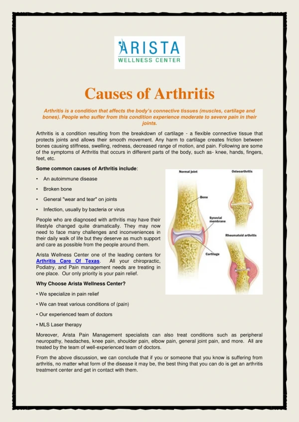
Arthritis of the Hands. On the Agenda. Normal Osteoarthitis Rheumatoid arthritis CPPD crystal deposition Gout Psoriatic arthritis. Normal Hand X-ray. Osteoarthritis (DJD). Gradual degeneration of articular cartilage Joint pain relieved with rest

E N D
On the Agenda • Normal • Osteoarthitis • Rheumatoid arthritis • CPPD crystal deposition • Gout • Psoriatic arthritis
Osteoarthritis (DJD) • Gradual degeneration of articular cartilage • Joint pain relieved with rest • Morning stiffness resolves within 30 minutes • Traditionally affects DIPs, 1st IP • No systemic symptoms • Painless nodules • Heberden’s at DIPs • Bouchard’s at PIPs
OA – Radiographic findings • Joint space narrowing • Osteophyte formation (white arrow) • Subchondral sclerosis (black arrows)
Joint space narrowing distally • Marginal osteophytes • Relatively unchanged proximal structures
Another example of OA • Oblique and AP views • 1st carpal metacarpal shows decreased joint space and subchondral sclerosis • 2nd and 3rd DIP shows osteophytes and subchondral sclerosis (Heberden’s nodes)
Rheumatoid Arthritis • Systemic inflammatory disease • Affects synovial membranes • Pannus (granulation tissue) develop in joint spaces and erode into the articular cartilage and bone • Prolonged morning stiffness (>1 hr) • PIPs, MCPs, and wrist commonly involved • Symmetric joint involvement
RA radiography - early • Earliest signs include soft tissue swelling due to effusion, tenosynovitis, and edema • Periarticular osteopenia • Marginal erosions often first seen at 2nd and 3rd MCPs and 3rd PIP articulations
Severe erosive changes at radio-ulnar joints carpal bones at the metacarpal heads • Bilaterally symmetric
Advanced RA • Boutonniere (top) • Swan neck • Labs: • +RF in 80%: IgM against Fc of IgG • Elevated ESR • Anemia of chronic disease
RA - Late • Complete MCP involvement • Large marginal erosions have nearly destroyed the joints • Bones are lucent due to osteopenia • Ulnar deviation
RA Bone Scan • Technetium-99 bone scan • Uptake shown in subclinical inflammation of joints • Symmetrical • Polyarticular
Calcium pyrophosphate dihydrate crystals (CPPD) • “Pseudogout” • Associated with metabolic diseases such as hyperparathroidism, hemochromatosis, hypothyroidism • Compared to gout: • Large joints affected (2nd to 5th MCPs, radio-carpal) • Rhomboid crystals • Positive birefringence • Calcification of articular cartilage • No cortical erosions
CPPD • Chondrocalcinosis • Distal radius and MCPs (2nd and 3rd) • Cartilage destruction similar to OA – differentiate by location • Location similar to RA – differentiate by absense of erosions • Calcium deposition at triangular fibrocartilage of the wrist (picture)
CPPD • Diffuse condrocalcinosis at the radiocarpal joint, the MCP joints and the PIP • Joint space narrowing, sclerosis, and subchondral cysts within the carpals
Gout • Disorder of purine metabolism – overproduction versus underexcreation • Deposition of urate crystals in joint spaces • Middle-aged men • Acute onset of extreme pain in small joints with redness and swelling • Needle shaped crystals with negative bifringence • Asymmetric, monoarticular
Gout Radiography • All joints of hand and wrist possible (2nd-5th PIP most common) • Soft tissue swelling • Well demarcated osseous erosions with sclerotic rims and overhanging edges • No decrease in bone density • Tophi not calcified • Relative sparing of joint space until late in the disease • Long latent period between onset of symptoms and radiographic changes
Psoriatic Arthritis • HLA-B27 positive, RF negative • Inflammatory • Seronegative spondyloarthropathy • Asymmetric and bilateral • Primarily distal involvement associated with nail changes • No periarticular osteoporosis • Five different patterns • Usually accompanies skin disease
Psoriatic Arthritis – Rad findings • Asymmetric proliferative erosions with ill-defined margins • Periosteal reaction • Soft tissue swelling • “Pencil-in-cup” deformity • Resorption of distal phalangeal tufts • Subluxation
Source • http://rad.usuhs.mil/medpix • Additional listed on request