Station 1
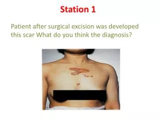
Station 1. Patient after surgical excision was developed this scar What do you think the diagnosis?. -Patient after surgical excision was developed this scar What do you think the diagnosis? It is a keloid ( cuz it’s beyond wound line and is itchy) -What is a keloid ?

Station 1
E N D
Presentation Transcript
Station 1 Patient after surgical excision was developed this scar What do you think the diagnosis?
-Patient after surgical excision was developed this scar What do you think the diagnosis? It is a keloid(cuz it’s beyond wound line and is itchy) -What is a keloid? is a type of scar beyond the site of wound. It is a result of an overgrowth of granulation tissue (collagen type 3) at the site of a healed skin injury which is then slowly replaced by collagen type 1
What is the difference between keloid and hypertrophic scar? • Hypertrophic scar: After the skin is injured, the healing process usually leaves a flat scar. Sometimes the scar is hypertrophic, or thickened, but confined to the margin of the wound
-Keloid: Keloids, by contrast, may start some time after the injury and extend beyond the wound site. What is the sign and symptoms of keloid? -raised and look shiny and dome-shaped -tend to be itchy, tender, or even painful to the touch. -ranging in color from pink to red. - causing potential cosmetic problems,
What is the cause? • Doctors do not understand exactly why keloids form in certain people or situations and not in others. Changes in the cellular signals that control growth and proliferation may be related to the process of keloid formation, • Location? can develop in any place that an abrasion has occurred, insect bites, scratching, burns, or other skin trauma.
Which people are most susceptible to keloids? • -Keloids are equally common in women and men, although at least in times past more women developed them because of a greater degree of earlobe and body piercing among women. • -In some cases, the tendency to form keloids seems to run in families.
Treatment: -silicone sheeting. -Surgery -Dressings — Moistened wound coverings made of silicone gel -Steroid injections — Steroid injections are best used as the scar begins to thicken or if the person is a known keloid former. Compression — Compression bandages applied to the site over several months, -Cryosurgery -Radiation therapy -Laser therapy
Station 2 Q. describe what you see Q. what is the most likely diagnosis ? Q. mention 2 features support your diagnosis Q. mention 3 differential diagnosis ?
Q1: describe what u see? • Small pigmented round superficial ulcer (can be nodule) in the right side blow the eye ,rolled edge, red brown in color ,base cover by”( depend on what u see >> dried serum and epithelium bleed easily) • دااااااااااائما اوصفوا اللي تشوفونه بالتفصيل >>>د.كردي • Q2: what is the most likely diagnosis ? • BCC • Q3:give us 2 features support your diagnosis? • In the face(above the line drawn from the mouth angel to the lob of ear) • In the espouser area to sun • Old age ( from picture) • Q4: give 3 dxdx? • Keratocnthosis ,m.Melanoma • Seq. cell ca ,syphilis
NB • BBC: • invasive locally not metastases • located on the face • nodular is the most common type • Rx surgery & radiation.(sensitive) • More in old age & it grow v. slowly
Note- melanoma : • Radio resistant. • Age 20_30. • Site ; lower limb in female , trunk in male . • Ssm most comm. Type
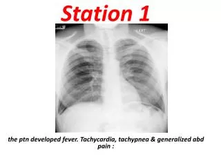
Station 3 Q. describe what you see Q. what is the most likely diagnosis ? Q. mention 2 features support your diagnosis Q. mention 3 differential diagnosis ?
Q1: describe what u see? Thin nodular lesion with an irregular edge & varying degree of pigmentation(pink, brown ,black) 2_3 cm ,oval In shape , some pigmentation in around . Q2: what is the most likely diagnosis ? Superficial spreading m. melanoma with an amelnotic nodular Q3:give us 2 features support your diagnosis? ABCD: asymmetry, border irregular, color variegation &diameter >6mm Q4: give 3 dxdx? Benign pigmented nevus. Benign hairy nevus. Jadassohn's nevus. Pigmented seborrheickeratosis Juvenile melanoma
ماحبيت اطول السلايدات بس هذي صور لكل انواع الميلانوما ان شاء الله يكون مفيد بالتوفيق كلنا يارب دعواتكم شيماء