Distortion
Distortion. By Prof. Jarek Stelmark. Distortion Distortion results from the radiographic misrepresentation of either the size (magnification) or shape of the anatomic part. When the image is distorted, recorded detail is also reduced. SIZE DISTORTION (MAGNIFICATION)


Distortion
E N D
Presentation Transcript
Distortion By Prof. Jarek Stelmark
Distortion Distortion results from the radiographic misrepresentation of either the size (magnification) or shape of the anatomic part. When the image is distorted, recorded detail is also reduced.
SIZE DISTORTION (MAGNIFICATION) The term size distortion/magnification refers to an increase in the object's image size compared with its true, or actual, size. Radiographic images of objects are always magnified in terms of the true object size. The distances used (SID and OID) play an important role in minimizing the amount of size distortion of the radiographic image.
As OID increases, size distortion (magnification) increases; as OID decreases, size distortion (magnification) decreases.
Because radiographers produce radiographs of three-dimensional objects, some size distortion always occurs as a result of OID. Even if the object is in close contact with the image receptor, some part of the object will be farther away from the image receptor than other parts of the object. Those parts of the object that are farther away from the image receptor will be represented radiographically with more size distortion than parts of the object that are closer to the image receptor.
Source-to-Image Receptor Distance SID also influences the total amount of size distortion represented on a radiograph. Although OID has the greatest effect on size distortion, SID is still an important factor for the radiographer to control in order to minimize size distortion. SID is inversely related to magnification.
As SID increases, size distortion (magnification) decreases; as SID decreases, size distortion (magnification) increases.
This is the reason that chest radiographs are obtained at a minimum SID of 72 inches (180 cm) rather than of 40 to 48 inches (100 to 122 cm), which is commonly used for most other examinations. A 72-inch (180 cm) SID results in less magnification of the heart and other structures within the thorax.
Minimum 40-inch (or 100-cm) SID It has been a long-standing common practice to use 40 inches (rounded to 100 cm) as the standard SID for most skeletal radiographic examinations. However, in the interest of improving image resolution by decreasing magnification and distortion, it is becoming more common to increase the standard SID to 44 or 48 inches (112 or 122 cm). Additionally, it has been shown that increasing the SID from 40 to 48 inches reduces the entrance or skin dose even when the requirement for increased mAs is considered. In this textbook, the suggested SID listed on each skeletal positioning page is a minimum of 40 inches, with 44 or 48 inches recommended if the equipment and departmental protocol allow.
Calculating Magnification To observe the effect of distance on size distortion, it is necessary to consider the magnification factor. The magnification factor (MF) indicates how much size distortion or magnification is demonstrated on a radiograph. The MF can be expressed mathematically by the following formula:
Determining Object Size On a PA chest film taken with an SID of 72 inches and an OID of 3 inches (SOD is equal to 69 inches), the size of a round lesion in the right lung measures 1.5 inches in diameter on the radiograph. The MF has been determined to be 1.044. What is the object size of this lesion?
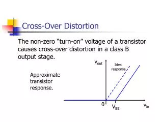
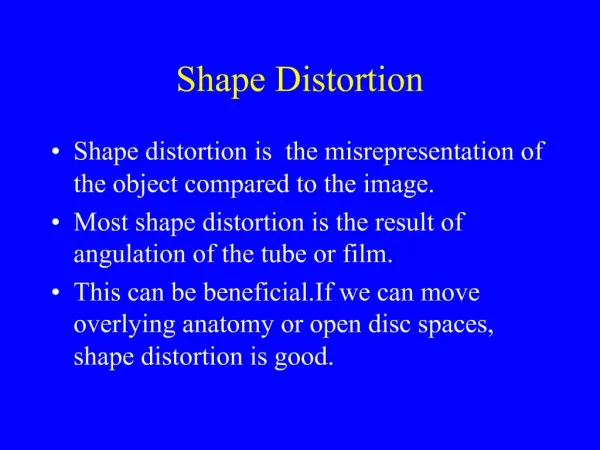
SHAPE DISTORTION In addition to size distortion, objects that are being imaged can also be misrepresented radiographically by distortion of their shape. Shape distortion can appear in two different ways radiographically: elongation or foreshortening. Elongation refers to images of objects that appear longer than the true objects. Foreshortening refers to images that appear shorter than the true objects.
Minimizing Shape Distortion • Elongation and foreshortening can be minimized by ensuring the proper CR alignment of the following: • X-ray tube • 2. Part • 3. Image receptor • 4. Entry or exit point of the CR
Sometimes, shape distortion is used to an advantage in particular projections or positions. CR angulation, for example, is sometimes required to elongate a part so that a particular anatomic structure can be visualized better. Also, CR angulation is sometimes required to eliminate superimposition of objects that normally would obstruct visualization of the area of interest. In general, shape distortion is not a necessary or desirable characteristic of radiographs.