Shape Distortion
Shape Distortion. Shape distortion is the misrepresentation of the object compared to the image. Most shape distortion is the result of angulation of the tube or film. This can be beneficial.If we can move overlying anatomy or open disc spaces, shape distortion is good. Shape Distortion.

Shape Distortion
E N D
Presentation Transcript
Shape Distortion • Shape distortion is the misrepresentation of the object compared to the image. • Most shape distortion is the result of angulation of the tube or film. • This can be beneficial.If we can move overlying anatomy or open disc spaces, shape distortion is good.
Shape Distortion • The ideal x-ray image would be to see the area of interest free of overlying structures. • A cephalad tube angle is toward the head. • A caudal tube angle is toward the feet. • The direction and amount of angulation is determined by the angle of the area of interest.
Tube Angle: Cephalad • Tube angled toward head. • Reduce SID one inch for every five degrees of tube angulation. • Used to get angled body parts perpendicular to film.
Cephalad Tube Angle • The amount of tube angle is determined by adding the angle to 90 for erect radiographs. • Anterior structures are on top of the image. • Posterior structures are projected to the bottom of the image.
Cephalad Tube Angle • This is an A-P Cervical Spine view. • The tube angle is 15° cephalad. • The tube is set to 105° • The central ray is parallel to the lower disc spaces.
Tube Angle: Caudal • Tube angled toward the feet. • Decrease SID by one inch for every five degrees of tube angulation. • Directs Beam through angled body part.
Caudal Tube Angle • The required tube angle is subtracted from 90 for erect radiography. • Posterior structures move to the top of the film. • Anterior structures move to the bottom.
Caudal Tube Angle • The patient is positioned A-P. • The tube is angled 15° caudal or set to 75° • This is the wrong tube angle to see the cervical spine.
Tube Angulation • Erect Radiography requires adding or subtracting the required angle from 90 degrees. • Cephalad angles: Add required angle to 90 degrees. • Caudal angles: Subtract required angle from 90 degrees.
Tube Angles • The direction will be affected by the angle of the anatomy. • Example: Sacral Base Angle 30-40° • A-P angle would be 30° cephalad • P-A angle would be 30° caudal.
No Tube Angle • If we wanted to see the top two cervical vertebra, no tube angle is needed. • If the mouth was open, we would see the dens well without a tube angle. • No shape distortion.
P-A Skull Film • The P-A or A-P skull is taken without any tube angle..
Chamberlain-Townes • The Townes Projection is part of a routine skull series. • The skull is in the same relative position as the P-A view • The tube is angled to throw the anterior part of the skull away from the occipital region of the skull.
Tube Angle Observations • 1. Which view has the mandible obscuring the A-P cervical spine? • Caudal tube angle • 2. If the mouth was open, which view would best demonstrate C-1 and C-2? • No tube angle
Tube Angle Observations • 3. Which view best demonstrates the cervical disc spaces? • Cephalad Tube Angle • 4. When the tube was set to 90° was there any tube angle in relation to the patient or film? • No
Tube Angle Observations • 5. When the tube was set at 105° was this a cephalad or caudal angle? • Cephalad • 6. What do we need to do to account for tube angles with erect radiography. • Add angle to 90° for cephalad angles. • Subtract angle from 90° for caudal angles.
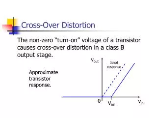
Beam Centering • The central ray must be centered to the area of interest . • If the beam is not centered, the shape of the area will be distorted.
Shape Distortion • The object of interest must be parallel to the film to avoid foreshortening. • If the object is angled in relation to the central ray, the object will have shape distortion.
Beam Centering • The center image has the beam properly aligned. • The right and left images have the beam not centered properly.
Observations for Beam Centering • 1. Which image most accurately demonstrated the true shape of the object? • With the beam centered to the object. • 2. When the tube is improperly centered too high or too low, what happens to the image. • The top structures are projected off center opposite to the direction of tube shift.
The End Return to Lecture Home Page