Connective
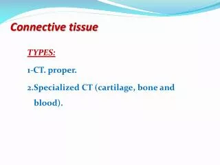
Connective. Loose (areolar, adipose, reticular) 2. Dense (dense irregular, white fibrous) 3. Cartilage (hyaline, elastic, fibrocartilage) 4. Bone 5. Blood 6. Mesenchyme Functions Support B. Connecting parts C. Storage (calcium, energy) D. Protection (skull, ribs, kidneys)

Connective
E N D
Presentation Transcript
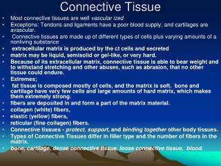
Connective Loose (areolar, adipose, reticular) 2. Dense (dense irregular, white fibrous) 3. Cartilage (hyaline, elastic, fibrocartilage) 4. Bone 5. Blood 6. Mesenchyme Functions Support B. Connecting parts C. Storage (calcium, energy) D. Protection (skull, ribs, kidneys) E. Movement (muscle attachments) F. Blood formation and transport 1.28
Thin, wire-like, flexible Common, long, white, wavy Short, branching (netting) CT Glue Chondroitin sulfate 1.30
Figure 4.7 Areolar connective tissue: A prototype (model) connective tissue. Cell types Extracellular matrix Ground substance Fibers • Collagen fiber • Elastic fiber • Reticular fiber Macrophage Fibroblast Cells? Blood vessels? Nerves? Lymphocyte Fat cell Capillary Mast cell Neutrophil Pg 128
Figure 4.13 Embryonic germ layers and the primary tissue types they produce. 16-day-old embryo (dorsal surface view) Muscle and connective tissue (mostly from mesoderm) Ectoderm Mesoderm Nervous tissue (from ectoderm) Endoderm Mesenchyme -ameboid Stem cells Flexible Epithelium Pg 144
Figure 4.8k Connective tissues. Liquid Plasma Water soluble fibers (k) Others: blood Description: Red and white blood cells in a fluid matrix (plasma). Plasma Function: Transport of respiratory gases, nutrients, wastes, and other substances. Neutrophil Location: Contained within blood vessels. Red blood cells Lymphocyte Photomicrograph: Smear of human blood (1860x); two white blood cells (neutrophil in upper left and lymphocyte in lower right) are seen surrounded by red blood cells. Pg 137
Figure 4.8a Connective tissues. Jello! Filler tissue (a) Connective tissue proper: loose connective tissue, areolar Description:Gel-like matrix with all three fiber types; cells: fibroblasts, macrophages, mast cells, and some white blood cells. Elastic fibers Function: Wraps and cushions organs; its macrophages phagocytize bacteria; plays important role in inflammation; holds and conveys tissue fluid. Collagen fibers Location: Widely distributed under epithelia of body, e.g., forms lamina propria of mucous membranes; packages organs; surrounds capillaries. Fibroblast nuclei Epithelium Photomicrograph: Areolar connective tissue, a soft packaging tissue of the body (300x). Lamina propria Pg 131
Figure 4.8b Connective tissues. (b) Connective tissue proper: loose connective tissue, adipose Description: Matrix as in areolar, but very sparse; closely packed adipocytes, or fat cells, have nucleus pushed to the side by large fat droplet. Function: Provides reserve food fuel; insulates against heat loss; supports and protects organs. Nucleus of fat cell Location: Under skin in the hypodermis; around kidneys and eyeballs; within abdomen; in breasts. Vacuole containing fat droplet Adipose tissue Triglycerides Photomicrograph: Adipose tissue from the subcutaneous layer under the skin (350x). Mammary glands Pg 131
Figure 4.8c Connective tissues. Supports soft organs (c) Connective tissue proper: loose connective tissue, reticular Description: Network of reticular fibers in a typical loose ground substance; reticular cells lie on the network. Function: Fibers form a soft internal skeleton (stroma) that supports other cell types including white blood cells, mast cells, and macrophages. White blood cell (lymphocyte) Location: Lymphoid organs (lymph nodes, bone marrow, and spleen). Reticular fibers Spleen Photomicrograph: Dark-staining network of reticular connective tissue fibers forming the internal skeleton of the spleen (350x). Pg 132
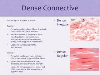
Figure 4.8e Connective tissues. Dermis, Deep fascia (e) Connective tissue proper: dense connective tissue, dense irregular Description: Primarily irregularly arranged collagen fibers; some elastic fibers; major cell type is the fibroblast. Nuclei of fibroblasts Function: Able to withstand tension exerted in many directions; provides structural strength. Location: Fibrous capsules of organs and of joints; dermis of the skin; submucosa of digestive tract. Collagen fibers Fibrous joint capsule Photomicrograph: Dense irregular connective tissue from the dermis of the skin (400x). Pg 133
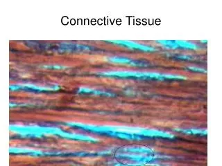
Figure 4.8d Connective tissues. (White Fibrous) (d) Connective tissue proper: dense connective tissue, dense regular Description: Primarily parallel collagen fibers; a few elastic fibers; major cell type is the fibroblast. Collagen fibers Function: Attaches muscles to bones or to muscles; attaches bones to bones; withstands great tensile stress when pulling force is applied in one direction. Location:Tendons, most ligaments, aponeuroses. Nuclei of fibroblasts Shoulder joint Ligament Photomicrograph: Dense regular connective tissue from a tendon (500x). Tendon Pg 133
Figure 4.8f Connective tissues. (f) Connective tissue proper: dense connective tissue, elastic Description: Dense regular connective tissue containing a high proportion of elastic fibers. Function: Allows recoil of tissue following stretching; maintains pulsatile flow of blood through arteries; aids passive recoil of lungs following inspiration. Elastic fibers Location:Walls of large arteries; within certain ligaments associated with the vertebral column; within the walls of the bronchial tubes. Aorta Photomicrograph: Elastic connective tissue in the wall of the aorta (250x). Heart Pg 134