Cell cycle control
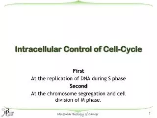
Cell cycle control. Cell division, role of cyclins and cyclin -dependent kinases (CDK) Structure, activation and deactivation of the cyclin -CDK complex Gene activation downstream of CDKs Cell cycle checkpoints Control of DNA integrity and the p53 protein

Cell cycle control
E N D
Presentation Transcript
Cell cycle control • Cell division, role of cyclins and cyclin-dependent kinases (CDK) • Structure, activation and deactivation of the cyclin-CDK complex • Gene activation downstream of CDKs • Cell cycle checkpoints • Control of DNA integrity and the p53 protein • DNA damages and repair mechanisms • Mutations, epi-mutations and cancer
Douglas Hanahan and Robert A. Weinberg (2000) The Hallmarks of Cancer. Cell 100 : 57–70 Cell division
Cellular cycles are generally NOT synchronous The cell cycle G0 Mitosis In the resting state (G0), cells do not divide Gap 2 • The distribution of cell at each stage of the cell cycle can be studied by flow cytometry and fluorescent DNA staining • Cell cycle synchronization • - Protein synthesis inhibitors • - Microtubule polymerization inhibitors (nocodazole) • - Knockout of cyclin-CDK complexes (for instance in thermosensitive mutants Gap 1 DNA Synthesis
Flow cytometry Fluorescence level Up to 8 fluorophores can be simultaneously analyzed Forward and side scattering is used to analyze cell size and granularity Analysis rate ≈ 10000 cells/sec Several analysis in parallel http://www.abcam.com/
The Fluorescence Activated Cell Sorter (FACS) To quantify DNA, a membrane-permeant fluorescent DNA marker is often used (Hoechst)
Synchronization of cell cycle • When cells proliferate, cyclins are continuously produced and degraded at specific times by the proteasome • When cells proliferate, cyclindependant kinases (CDK) are expressed. CDK alone are inactive, cyclin-CDK complexes are progressively active (see later) • Different Cyclin-CDK complexes form during the cell cycle • In yeast, two complexes exist : the Start Promoting Factor at the G1/S transition and the M-phase Promoting Factor during mitosis • In vertebrates, five cyclin-CDK complexes form during the cell cycle • Cyclin-CDK complexes are regulated by phosphorylation, proteolytic cleavage and partial degradation in the proteasome • Cyclin-CDK complexes have specific kinase activity
Cell cycle regulation by cyclin-dependent kinases phospho-rylation of specific targets phosphorylation of specific targets phospho-rylation of specific targets phosphorylation of specific targets
H NH C CO H CH3 C OH H H H NH C CO HN C CO H H C NH C CO CH2 O H H C OH O OH P O- OH Protein phosphorylation Threonine Thr, T Phosphorylated serine Ser-P, S-P Serine Ser, S kinase + ATP + ADP + H+ Tyrosine Tyr, Y • Serine (S), Threonine (T) and Tyrosine (Y) are the main amino acids that can be phosphorylated. Kinase specificity is determined by the surrounding sequence (consensus sequence) • Phosphorylation modifies the H bonding network around the modified amino acid. • Phosphorylation adds a negative charge to the modified amino acid. • Phosphorylation can be mimicked by mutation to a negatively charged amino acid, such as aspartate (D) or glutamate (E). • Phosphorylation can be prevented by mutation to an amino acid lacking the hydroxyl group : S/T A, Y F
H H NH C CO H H C NH C CO O H H C OH O OH P O- Serine Ser, S Phosphorylated serine Ser-P, S-P kinase - - - - -
Example : structure of the cyclin-dependent kinase – cyclin complex cyclin ATP Substrate binding site : X-P-Xaa-(S/T)-P-X where Xaa is a neutral of basic amino acid from PDB 2CCH cyclin-dependant kinase
Serine Ser, S H H NH C CO NH C CO H H C H H C O OH O OH P O- Protein dephosphorylation Phosphorylated serine Ser-P, S-P + H2PO4− H2O + phosphatase • 518 kinases and 73 phosphatases have been described in the human genome • Phosphorylation or dephosphorylation of specific aminoacids changes the conformation of proteins or allows their binding to other partners • Once phosphorylated, the phosphorylated protein dissociates from the kinase • Once dephosphorylated, the dephosphorylated protein dissociates from the phosphatase
Consequences of protein phosphorylation protein kinase phosphatase Phosphorylated protein Conformational change New partner binding Activation/Inactivation/ Degradation
Example : inactivation of Src, a soluble tyrosine-kinase Regulatory domain Phospho-tyrosine polyproline Polyproline binding site Catalytical domain tyrosine ATP phosphorylation Active kinase Inactive kinase dephosphorylation
The human kinome • 518 kinases with specificity towards serine, threonine, tyrosine and histidine residues have been described • Kinases regulate a plethora of cellular processes in all organisms • Serine/Threonine kinases constitute the main fraction of the kinome, they are predominantly involved in regulating signaling processes • Tyrosine kinases represent a small fraction of the kinome (58 RTK, 32 CTK) and only exist in eukaryotes, they are predominantly involved in regulating signaling processes. Gibbins,J.M. (2004). Study of tyrosine kinases and protein tyrosine phosphorylation. Methods Mol. Biol. 273, 153-16. Ostman,A. and Bohmer,F.D. (2001). Regulation of receptor tyrosine kinase signaling by protein tyrosine phosphatases. Trends Cell Biol. 11, 258-26. Hubbard,S.R. and Till,J.H. (2000). Protein tyrosine kinase structure and function. Annu. Rev. Biochem. 69, 373-398. Manning,G., Whyte,D.B., Martinez,R., Hunter,T., and Sudarsanam,S. (2002). The protein kinase complement of the human genome. Science 298, 1912-1934. http://www.nih.go.jp/mirror/Kinases/ http://www.manros-therapeutics.com/artwork/science/human-kinome
Activation of the cyclin-dependent kinase – cyclin complex cyclin Inhibitory phosphorylation site : Y15 ATP Substrate binding site : X-P-Xaa-(S/T)-P-X where Xaa is a neutral of basic amino acid Activatory phosphorylation site : T161 cyclin-dependent kinase from PDB 2CCH
CDK kinase structure and activation dominant mutant Y15F Inhibitory phosphorylation site : Y15 Cyclin interaction domain Regulatory loop : T-loop null mutant T161A Activatory phosphorylation site : T161 Catalytic domain T161-phos • Inaccessible substrate binding site • Substrate binding site exposed
Control of the cyclin-CDK complex activity Inactive cyclin-CDK complex Inactive cyclin-CDK complex CAK : CDK CDK inhibitory Y15 Cyclin activatory T161 - phosphatase Active cyclin-CDK complex degradation + protease Cyclin + P Example of yeast mitosis promoting factor
A positive feedback loop in the activation of the cyclin-CDK complex + cdk + cyclin Inactive cyclin-CDK Active cyclin-CDK + Dt cdk • Activation of cyclin-CDK is self sustained activation threshold • Activation of cyclin-CDK triggers its own deactivation delay cyclin Active cyclin-CDK threshold time delay
Activation time 10 msec Positive feedback close to the threshold speeds up the propagation of chemical signals + cdk + cyclin Inactive cyclin-CDK Active cyclin-CDK + Dt Average distance betweenmolecules at 1 µM : 100 nm cdk distance iBio-SeminarsDAVID MORGAN: controlling the cell cycle, Anaphase onset:
Cyclin structure and inactivation proteasome ubiquitinylation Destruction box 10 20 30 40 50 60 MALRVTRNSK INAENKAKIN MAGAKRVPTA PAATSKPGLRPRTALGDIGN KVSEQLQAKM 70 80 90 100 110 120 PMKKEAKPSA TGKVIDKKLP KPLEKVPMLV PVPVSEPVPE PEPEPEPEPV KEEKLSPEPI 130 140 150 160 170 180 LVDTASPSPM ETSGCAPAEE DLCQAFSDVI LAVNDVDAED GADPNLCSEY VKDIYAYLRQ 190 200 210 220 230 240 LEEEQAVRPK YLLGREVTGN MRAILIDWLV QVQMKFRLLQ ETMYMTVSII DRFMQNNCVP 250 260 270 280 290 300 KKMLQLVGVT AMFIASKYEE MYPPEIGDFA FVTDNTYTKH QIRQMEMKIL RALNFGLGRP 310 320 330 340 350 360 LPLHFLRRAS KIGEVDVEQH TLAKYLMELT MLDYDMVHFP PSQIAAGAFC LALKILDNGE 370 380 390 WTVKNKYATS KHAKISTLPQ LNSALVQDLA KAVAKV Human cyclin B1
(Thermosensitive) mutants of the yeast cell cycle and discovery of Cyclin Dependent Kinases (CDK) Cdc: Cell division cycle Saccharomyces pombe : about 70 cdc genes (in 6800) L.H. Hartwell et al. 1960-70 Cdc2 = Cdk1 Hyperactive mutant Null mutant wee1- cdc25-
Cyclin-CDK activity Discovery of cyclins Histone phosphorylation • Cyclins accumulate during the cell cycle and are degraded after mitosis (Tim Hunt 1981) • Cyclins are the only proteins whose synthesis is required to drive the cell cycle (Mark Kirschner) Marie-Anne Felix, Jonathon Pines, Tim Hunt and Eric Karsenti, 1989, EMBO Journal 8 : 3059-3069 Active Cyclin-CDK
Cell cycle synchronization A transcriptome view of yeast cell cycle Saccharomyces cerevisiae: 800 genes are regulated during the cell cycle (out of 6800) green: over-expression compared to the average level red: under-expression compared to the average level Genes that are regulated during the cell cycle are ordered as a function of their up-regulation during the cell cycle Spellman et al. 1998 Mol. Biol. Cell time
MCM : genes regulated as the transcription factor Mcm1 (G1) CLB2 : genes regulated as cyclin B Clb2 (G2-M) SIC1 : genes regulated as Sic1, the inhibitor the of the Clb2-Cdc28 complex CLN2 : genes regulated as Cln2 (G1-S) Histones : proteins associated to DNA in nucleosomes Synchronisation du cycle cellulaire Set of co-regulated genes during the cell cycle Co-regulated genes have similar promoter sequences (and probably common transcription factors) Example : MCM consensus promoter
+ + ADP ADP ATP ATP TRANSCRIPTION of S-phase genes Activation of the E2F transcription factor cyclin D1 cyclin E P P cdk4 cdk2 P P P P Rb Rb* Rb* P P - Rb : retinoblastoma gene RNA pol E2F E2F : transcription factor (E2 gene of adenovirus) E2F binding site G1 phase S phase
P P P P Rb* Rb + + + ADP ADP ADP P P ATP ATP ATP P P P P DP DP Deactivation of the E2F transcription factor cyclin D1 cyclin A cyclin E P P P cdk4 cdk2 cdk2 DP Rb* Rb : retinoblastoma gene - DP : (hetero) dimerization protein RNA pol E2F E2F : transcription factor (E2 gene of adenovirus) E2F binding site G1 phase S phase
Cell cycle control • Cell division and the discovery of cyclins and cyclin-dependent kinases (CDK) • Structure, activation and deactivation of the cyclin-CDK complex • Gene activation downstream of CDKs • Cell cycle checkpoints • Control of DNA integrity and the p53 protein • DNA damage and repair mechanisms • Mutations, epimutations and cancers
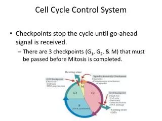
An overview of the cell-cycle control system The core of the cell-cycle control system consists of a series of cyclin-Cdk complexes(yellow). The activity of each complex is also influenced by various inhibitory checkpoint mechanisms, which provide information about the extracellular environment, cell damage, and incomplete cell-cycle events (top). These mechanisms are not present in all cell types; many are missing in early embryonic cell cycles, for example.
p53 Retinoblastoma protein (Rb) Anaphase Promoting Complex (APC) APOPTOSIS APOPTOSIS APOPTOSIS Cell cycle checkpoints
The p53 protein holds the cell cycle at the G1/S checkpoint in the presence of DNA damage • p53 is a tetrameric 393 aa protein • p53 consists of 3 domains : 1 100 200 300 393 phosphorylations NLS acetylations transcription activation domain DNA binding domain regulatory domain • The transcription activation domain interacts with the Mdm2 protein that triggers p53 degradation. • The DNA binding domain interacts with a specific DNA sequence that controls p21CIP expression • The conformation and the localization of p53 is controlled by phosphorylation and acetylation p53 DNA binding domains in complex with DNA
DNA intact damaged Chk inactive active p53 absent DNA bound (mdm2) (phosphorylated) p21 repressed expressed CDK active inactive Cycle G1S G1 blockage + Chk1/2 p21 + RNA polymerase p53-P Double strand breaks TRANSCRIPTION of p21, a CDK inhibitor Single strand breads (30 to 40 missing bases) cyclin E Basepair mismatches P cdk2 Mdm2 p53 p53-Ub synthesis degradation Mdm2 = murine double minute oncogene Chk = checkpoint kinase p21= CIP (cdk2 inhibiting protein) = WAF1 (Wild Type p53-activated fragment)
The p53 protein is mutated in 50% of human cancers Arg175, 249, 273, 282, Gly245 Arg248 Mutation frequency p53 primary sequence • These mutations decrease the affinity of p53 for DNA, which eliminates the G1/S restriction point controlled by the Cdk2-cyclinE complex
The link between p53 and apoptosis is complex • P53 is a transcription factor that controls multiple gene expression : it recognizes the sequence 5’-PuPuPuC(A/T)(A/T)GPyPyPy-3’ separated by 0-13 bp • P53 stops the cell cycle thanks to p21WAF1, GADD45 and 14-3-3s • P53-dependent apoptosis involves three groups of genes : • cell surface proteins : Fas (CD95), Killer/DR5, PERP that are death receptors activated by specific ligands • mitochondrial proteins : Bax, Bcl2, Bcl-XL, Bid, NOXA, PUMA, P53AIP… involved in mitochondrial inactivation during apoptosis • cytoplasmic proteins : PTEN that modulate extracellular signals • P53 regulated gene expression pattern differs in different cell types and in response to different signals • The expression of the target genes depend on other signals • When exposed to ionizing radiations, some cells undergo apoptosis and other arrest the cell cycle • In general, cells that are highly proliferating and relatively undifferentiated respond to g-irradiation by P53-mediated apoptosis; cells that divide slowly only stop dividing in a P53 dependent manner.
Extrinsic pathway P53-mediated apoptosis Intrinsic pathway
Cell cycle control • Cell division and the discovery of cyclins and cyclin-dependent kinases (CDK) • Structure, activation and deactivation of the cyclin-CDK complex • Gene activation downstream of CDKs • Cell cycle checkpoints • Control of DNA integrity and the p53 protein • DNA damage and repair mechanisms • Mutations, epimutations and cancers
Wood et al. (2007) The Genomic Landscapes of Human Breast and Colorectal Cancers Science 318 : 1108-1113 Cancer genomelandscapes PIK3CA FBXW7 P53 PIK3CA P53 RAS APC Nonsilentsomatic mutations are plotted in two-dimensional space representing chromosomal positions of RefSeq genes. The telomere of the short arm of chromosome 1 is represented in the rear left corner of the green plane and ascending chromosomal positions continue in the direction of the arrow. Chromosomal positions that follow the front edge of the plane are continued at the back edge of the plane of the adjacent row, and chromosomes are appended end to end. Peaks indicate the 60 highest-ranking CAN-genes for each tumor type, with peak heights reflecting CaMP scores (7). The dots represent genes that were somatically mutated in the individual colorectal (Mx38) (A) or breast tumor (B3C) (B) displayed. The dots corresponding to mutated genes that coincided with hills or mountains are black with white rims; the remaining dots are white with red rims. The mountain on the right of both landscapes represents TP53 (chromosome 17), and the other mountain shared by both breast and colorectal cancers is PIK3CA (upper left, chromosome 3). FBXW7 binds to cyclin E and probably targets cyclin E for ubiquitin-mediated degradation
DNA damages and epigenetic changes • 1. DNA damage : change in the DNA structure which is not replicated when the DNA is replicated : • Chemical modification of a base : adduct or disruption • Replacement of a base by another one : mismatch • Single or double strand break • Repair or permanent mutations • 2. Epigenetic modifications govern DNA accessibility and play an important role in gene regulation. They are transmitted to daughter cells during cell division. Epigenetic modifications act on : • DNA : covalent modification of DNA, for instance methylation of CpG islands repression • Histones : phosphorylation, methylations and acetylation of specific amino acids affect the affinity of DNA binding • Epigenetic changes (epimutations) affect gene expression • Epigenetic changes are potentially reversible
Origin of DNA damages : endogenous Table 1. DNA damages due to natural endogenous causes in mammalian cells Accurate repair mechanisms exist for all these damages, Bernstein et al. (2013) DNA Damage, DNA Repair and Cancer. INTEK chapter 16, p 413-465
DNA repair mechanisms Damage type Repair mechanisms Repair system • Recognition Nucleotide Excision Repair (NER) Base Excision Repair (including PARP enzymes) (BER) Mis-Match Repair (MMR) Base mismatch T-T dimers Adducts Single strand breaks • Restriction • Excision • Synthesis • Ligation Double strand breaks HomologousRecombinationalRepair (HRR) • Excision • Recombination • Ligation Non-homologous End Joining (NHEJ) • or direct ligation
Main DNA repair genes (169) Number of genes 21 8 30 20 11 16 3 17 8 11 14 2 3 5 2 9 HomologousRecombinationalRepair (HRR)Non-homologous End Joining (NHEJ) Nucleotide Excision Repair (NER) Base Excision Repair (including PARP enzymes) (BER) Mis-Match Repair (MMR) Fanconi Anemia (FANC) [affects HRR (above) and translesion synthesis (TLS)] Direct reversal of damage DNA polymerases (act in various pathways) Editing and processing nucleases (act in various pathways) Ubiquitination and modification/Rad6 pathway including TLS DNA damage response Modulation of nucleotide pools Chromatin structure Defective in diseases and syndromes DNA-topoisomerase crosslinks Other genes Table 5. DNA repair pathways and other processes affecting DNA repair [67, 68] Bernstein et al. (2013) DNA Damage, DNA Repair and Cancer. INTEK chapter 16, p 413-465
DNA repair mechanisms and cancer • 30% of cancers are familial (they correspond to mutations in predisposition genes) : many of these genes concern DNA repair mechanisms • Men have higher cancer incidence rates as women (unexplained) • Recessively transmitted genes • Both alleles are affected • DNA defects start as soon as the gene is expressed • e.g. ataxia telangiectasia Dominantly transmitted genes One allele is affected Two possibilities : 1. The mutated allele is dominant DNA defects start as soon as the gene is expressed e.g. certain truncated APC genes 2. The mutated allele is recessive A second inactivating mutation is necessary for the phenotype to appear e.g. most APC mutations requires a biallelic loss of APC function (“loss of heterozygosity” at the APC locus)
Tumor suppressors are genes that are able to induce cancer when both alleles are inactivated (cell transformation) checkpoints Cell signaling DNA repair enzymes Dominantly inherited cancer susceptibility
Inherited recessive DNA mutations that increase the risk of cancer checkpoints DNA repair enzymes Recessively inherited cancer susceptibility
DNA damages and life habits • 70% of cancers are considered to be sporadic • Evidence for environmental causes of colon cancer : • Black native africans : incidence rate 1 out of 100 000 • Male black africanamericans : incidence rate 63 per 100 000 • Other US male citizens : incidence rate 47 to 52 depending on ethnic group • Japanese in Miyagi vs Japanese in Hawaii (Immigration from Japan to Hawaii during 1868–1924
Origin of DNA damages : exogenous • The main causes of increased cancer incidence rates are : • Tobacco use • Diets higher in saturated fats bile acid oversecretion reactive oxygen species • Other exogenous DNA damaging agents are : Bernstein et al. (2013) DNA Damage, DNA Repair and Cancer. INTEK chapter 16, p 413-465
Exogenous DNA damaging agents in tobacco smoke Cunningham et al. (2011) A novel application of the Margin of Exposure approach: Segregation of tobacco smoke toxicants. Food and Chemical Toxicology 49 : 2921–2933 • Margin of Exposure : MOE = BMDL10/estimated daily human intake • BMDL10 or benchmark dose : the daily dose that causes a 10% increase in tumourincidence above control values in animal studies • Estimated daily human intake : corresponding to 20 cigarettes per day, assuming 100% retention of the constituent in the smoker’s body (average smoker consumption in France 14 cigarettes/day)
Acrolein adducts • Acrolein (2-propenal) is ubiquitously present in cooked foods and in the environment. It is formed from carbohydrates, vegetable oils and animal fats, amino acids during heating of foods, and by combustion of petroleum fuels and biodiesel. • Smoking of tobacco products equals or exceeds the total human exposure to acrolein from all other sources. • Acrolein forms adducts with DNA which are mutagenic and induce predominantly G:C-to-T:A transversion Jan F. Stevens and Meier (2008) Acrolein: sources, metabolism, and biomolecular interactions relevant to human health and disease. Mol Nutr Food Res. 52 : 7–25 Feng et al. (2006) Acroleinis a major cigarette-related lung cancer agent: Preferential binding at p53 mutational hotspots and inhibition of DNA repair. PNAS 103 : 15404–15409
translocation to the nucleus aromatic molecule (L) Aryl hydrocarbon Receptor AhR AhR-L AhR-L AhRE AhRE induction of specific mRNA (AhRE) P450 cytochromes (phase I) : CYP1A1, CYP1A2, CYP1B1, CYP2S1 Phase II enzymes : GST, UGT (detoxification mechanism) Growth Differentiation Metabolism (toxicity) CYP1A1, CYP1A2 epoxide hydrolase the diol epoxidecovalentlybinds to DNA (adductpoorlyrepaired) Increased DNA mutations (G:C to T:A transversions) & cancer benzo[a]pyrene-7,8-dihydrodiol -9,10-epoxide Metabolism et carcinogenicity of Benzo[a]Pyrene Benzo[a]pyrene is a product of incomplete combustion at temperatures between 300 and 600 °C. benzo[a]pyrene (BP)
Mutational spectrum of the human p53 gene in human lung cancers of cigarette smokers and nonsmokers (International Agency for Research on Cancer p53 Mutation Database, http:<949><949>www-p53.iarc.fr). Red bars represent mutations occurring at codons with CpG sequences, and black bars represent mutations occurring at codons without CpG sequences. Val 157 Arg 158, 175, 248, 249, 273, 282 Gly 245 Feng et al. (2006) PNAS 103 : 15404–15409