
Growth in Bone Length
E N D
Presentation Transcript
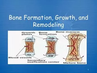
Growth in Bone Length • Growth in length occurs at the epiphyseal plate • Involves the formation of new cartilage by • Interstitial cartilage growth • Appositional growth on the surface of the cartilage • Closure of epiphyseal plate: epiphyseal plate is ossified becoming the epiphyseal line. Between 12 and 25 years of age • Articular cartilage: does not ossify, and persists through life • Appositional growth only • Interstitial growth cannot occur because matrix is solid • Occurs on old bone and/or on cartilage surface ex: trabeculae grow in size by the deposition of new bone matrix by osteoblasts onto the surface of the trabeculae.
Growth at Articular Cartilage • Increases size of bones with no epiphyses: e.g., short bones • Chondrocytes near the surface of the articular cartilage similar to those in zone of resting cartilage • The process of growth in articular cartilage is similar to that occurring in the epiphyseal plate, except that the chondrocyte columns are not as obvious. • When epiphyses reach their full size, the growth of cartilage & its replacement by bone cease. • However, articular cartilage persists throughout life & does not become ossified as does epiphyseal plate.
Factors Affecting Bone Growth • Size and shape of a bone determined genetically but can be modified and influenced by nutrition and hormones • Nutrition • Lack of calcium, protein and other nutrients during growth and development can cause bones to be small • Vitamin D • Necessary for absorption of calcium from intestines • Can be eaten or manufactured in the body • Rickets: lack of vitamin D during childhood (bowed bones) • Osteomalacia: lack of vitamin D during adulthood leading to softening of bones • Vitamin C • Necessary for collagen synthesis by osteoblasts • Scurvy: deficiency of vitamin C (causes ulceration & hemorrhage in body) • Lack of vitamin C also causes wounds not to heal (because requires collagen synthesis), teeth to fall out (because ligaments that hold them in place break down)
Factors Affecting Bone Growth (cont.) • Hormones • Growth hormone from anterior pituitary. Stimulates interstitial cartilage growth and appositional bone growth • Thyroid hormone required for growth of all tissues • Sex hormones such as estrogen and testosterone • Cause growth at puberty, but also cause closure of the epiphyseal plates and the cessation of growth
Bone Remodeling • Converts woven bone into lamellar bone • Caused by migration of Basic Multicellular Units (BMU) • Groups of osteoclasts and osteoblasts that remodel bones • Involved in bone growth, changes in bone shape, adjustments in bone due to stress, bone repair, and Ca ion regulation • Relative thickness of bone changes as bone grows. Bone constantly removed by osteoclasts and new bone formed by osteoblasts. • Formation of new osteons in compact bone • Osteoclasts enter the osteon from blood in the central canal and internally remove lamellae. Osteoblasts replace bone • Osteoclasts remove bone from the exterior and the bone is rebuilt
Bone Repair • Hematoma formation. Localized mass of blood released from blood vessels (damaged from fracture) but confined within an organ or space. Clot formation (consists of fibrous proteins that stop the bleeding). • Callus formation. Callus: mass of tissue that forms at a fracture site and connects the broken ends of the bone. - Internal- blood vessels grow into clot in hematoma (several days after fracture). • Macrophages clean up debris, osteoclasts break down dead tissue, fibroblasts produce collagen and granulation tissue. • Chondroblasts from osteochondral progenitor cells of periosteum and endosteum produce cartilage within the collagen. • Osteoblasts invade. New bone is formed. - External- collar around opposing ends. Periosteal osteochondral progenitor cells osteoblasts and chondroblasts. Bone/cartilage collar stabilizes two pieces.
Bone Repair, cont. • Callusossification. Callus replaced by woven, cancellous bone through endochondral ossification. • Bone remodeling. Replacement of cancellous bone and damaged material by compact bone. Sculpting of site by osteoclasts
Calcium Homeostasis • Bone is major storage site for calcium • The level of calcium in the blood depends upon movement of calcium into or out of bone. • Calcium enters bone when osteoblasts create new bone; calcium leaves bone when osteoclasts break down bone • Two hormones control blood calcium levels- parathyroid hormone (produced by parathyroid gland----blood calcium decreases then secretion of PTH increases which increases # of osteoclasts) and calcitonin ( produced by thyroid gland----decreases osteoclast activity
Effects of Aging on Skeletal System • Bone matrix decreases. More brittle due to lack of collagen; but also less hydroxyapatite. • Bone mass decreases. Highest around 30. Men denser due to testosterone and greater weight. African Americans and Hispanics have higher bone masses than Caucasians and Asians. Rate of bone loss increases 10 fold after menopause. Cancellous bone lost first, then compact. • Increased bone fractures • Bone loss causes deformity, loss of height, pain, stiffness • Stooped posture • Loss of teeth
Bone Fractures • Open (compound)- bone break with open wound. Bone may be sticking out of wound. • Closed (simple)- Skin not perforated. • Incomplete- doesn’t extend across the bone. Complete- does • Greenstick: incomplete fracture that occurs on the convex side of the curve of a bone • Hairline: incomplete where two sections of bone do not separate. Common in skull fractures • Comminuted fractures: complete with break into more than two pieces
Bone Fractures, cont. • Impacted fractures: one fragment is driven into the cancellous portion of the other fragment. • Classified on basis of direction of fracture • Linear • Transverse • Spiral • Oblique • Dentate: rough, toothed, broken ends • Stellate radiating out from a central point.