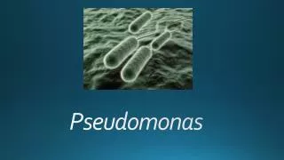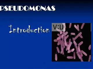
Pseudomonas
Pseudomonas. - Microscopic appearance. - Cultural characteristics. - Biochemical Tests of Pseudomonas species. - Identification of Pseudomonas using API 20E. Pseudomonas putida. Pseudomonas fluorescens. Pseudomonas aeruginosa. Pseudomonas aeruginosa causing :-.

Pseudomonas
E N D
Presentation Transcript
Pseudomonas - Microscopic appearance - Cultural characteristics - Biochemical Tests of Pseudomonas species. - Identification of Pseudomonas using API 20E
Pseudomonas putida Pseudomonas fluorescens
Pseudomonas aeruginosa causing :- - Skin infections ( wound swab )* *Specimens depending on the site of infection.
- Urinary Tract infection ( urine )* - Respiratory infections ( sputum & effusions )* - OtitisExterna ( ear swab )* *Specimens depending on the site of infection.
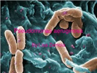
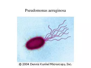
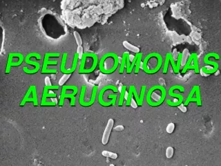
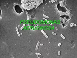
Pseudomonas aeruginosa is a Gram negative, non-sporing motile rod
P. aeruginosaproduces large, flat, spreading colonies which are often haemolytic and usually pigment-producing. The pigments diffuse into the medium giving it a dark greenish-bluecolour
P. aeruginosaproduces pale colouredcolonies on MacConkey agar
On Nutrient agar P. aeruginosacan be recognized by the pigments, it produces a blue-green pigment (pyocyanin).
O–F Test The oxidative-fermentative test determines if certain gram-negative rods metabolize glucose by fermentation or aerobic respiration (oxidation). During the anaerobic process of glucose fermentation, the high concentration of acid produced will turn the bromthymol blue indicator in OF media from green to yellow in the presence or absence of oxygen
Acid production in both the open and oil-covered tubes indicates a fermentative result ( e.g. Escherichia coli )
Acid production in the open tube and not the oil-covered tube indicates an oxidative result. P. aeruginosa incubated for 24 hours. P. aeruginosa incubated for 48 hours. P. aeruginosa incubated for 5 days.
No color change in the oil-covered tube and color change to alkaline in the open tube indicates a negative result. A. faecalis cannot use glucose fermentatively or oxidatively. The blue at the top of the open tube is due to amine production resulting from the metabolism of protein in the media.
Vibrio - Microscopic appearance - Cultural characteristics • Identification of Vibriocholeraebiochemically and • serologically
Vibriocholerae Vibrioalginolyticus Vibriovulnificus Vibrioparahaemolyticus
V. cholerae causes cholera • ( rice watery diarrhoea)* • V. parahaemolyticus causes • severe acute gastroenteritis( faeces)* • V. alginolyticus causes wound infection • ( wound swab )* • V. vulnificus causes wound infection and septicaemia • ( wound swab )*
Alkaline Peptone Water On Thiosulphate-citrate bile salt sucrose (TCBS) agar V. cholerae 01 and 0139 produce 2–3 mm in diameter sucrose-fermenting yellow colonies after overnight incubation at 35–37 ºC.
On TCBSV. mimicus, V. vulnificus and V. parahaemolyticusproduce green-blue non-sucrose fermenting colonies
V. cholerae 01 and 0139 grow on blood agar, (subcultured from alkaline peptone water), producing betahaemolytic colonies.
On MacConkey agar V. cholerae produces small non-lactose fermenting colonies.
Fermentation of L-arabinose. This test is of value in differentiating V. cholerae from V. fluvialis (both produce yellow colonies on TCBS agar). V. fluvialis ferments L-arabinose
SerotypingV. cholerae 01 and 0139:- Separate antisera are required to identify V. cholerae 01 (Inaba and Ogawa) and V. cholerae 0139 (Bengal). Isolates from TCBS cultures require subculturing to nutrient agar before carrying out serotyping.
In most countries V. cholerae 01 cholera is caused by the El Tor biotype. In India and Bangladesh where the Classical biotype also occurs, the Voges-Proskauer (VP) test can be used to differentiate the two biotypes. The El Tor biotype is VP positive and the Classicalbiotype is VP negative. When required other tests (haemagglutination and sensitivity to 50 iupolymyxin B) El Tor gives a positive agglutination test and is resistant to 50 iupolymyxin B. while Classicalbiotypes give a negative agglutination test and are sensitive to 50 iu polymyxin B.