Download

1 / 10
100 likes | 119 Vues
This project tackles the automated processing of telomeres in chromosomes for medical research, focusing on counting visible telomeres and analyzing telomere length using fluorescence probes. The detailed workflow includes image preprocessing, nuclei and telomere segmentation, and interactive editing of invalid nuclei. Complete CSV files are generated for statistical analysis in R, enabling comprehensive data summaries and graphical comparisons of telomere parameters across different illnesses.

E N D
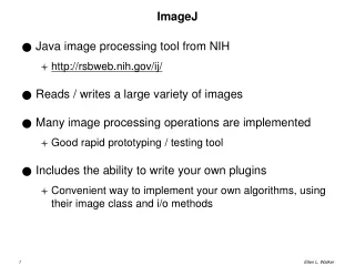
Automated processing of telomeres with ImageJ and R 5th ImageJ User & Developer Conference 3 September 2015 Aryeh Weiss Bar Ilan University
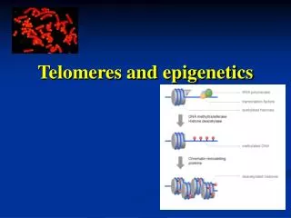
Telomere Imaging -- Problem Statement • Telomeres are regions of repetitive nucleotide sequences located at the ends of chromosomes. They are sub-resolution objects • Telomere length in various illnesses (particularly cancer) is a topic of research • We are interested in visible telomere count (per cell), and relative telomere length as indicated by the intensity of the emission of telomeres labeled with a fluorescent hybridization probe (PNA-Cy3).
Problem Statement -- continued • Each field includes 3-8 cells. • 20-40 fields are acquired from each slide. • 4-5 slides are prepared for each illness. • 4 illness + a healthy control were studied. • There are about 30-120 visible telomeres in each cells • If we do the math about 3000 cells will be studied, and about 200,000 telomeres will be counted.
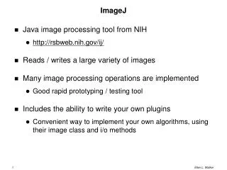
Image Processing • Image Processing Workflow: • Preprocess: remove point noise (median filter). Darknoise and nonuniform illumination correction. • Segmentation of nuclei (DAPI) • Creation of best focus image (max intensity projection) • Segmentation of telomeres • Editing of invalid nuclei. • Creation of telomere and cell CSV files (Python) • Statistical analysis in R
Acquisition of Z-stacks The telomeres are in focus in different planes of the z-stack 17 images were acquired at 0.2μm spacing. Above Middle Below
Segmentation of Nuclei • Intensity based segmentation. • Watershed algorithm to separate overlapping or touching nuclei • Shape based selection (roundness) • Output – a complete ROI (region of interest) list identifying each nucleus The metaphase spread was not marked because it fails the roundness criterion
Segmentation of Telomeres • Based on the “Find maxima” Imagej Plugin. • The plugin counts the telomeres in each cell, individually. • Mean intensity for each telomere is recorded • Output – a complete ROI (region of interest) list identifying each telomere in each cell (one CSV file/cell)
Interactive Editing of Invalid Nuclei Object 36 was removed because of its irregular shape. Object 37 was removed because of the overlap with the cell(s) below. Rules of the Game • All steps are saved – all edited nuclei are available for review • Nuclei that overlap with others may produce invalid counts • Nuclei that are irregular have probably been “dragged” along the glass and are removed.
Hierarchy of Data • Each cell CSV file with telomere intensities and locations • Each slide CSV file with one row/cell (telomere data summarized) • Each illness Multiple slides/illness combined and then analyzed in R • R output Charts comparing selected parameters (mean intensity, telomere count, percent “bright” telomeres…) in a graphical display.
Acknowledgements DrAlizaAmiel, Meir Hospital in KfarSaba Hila Katz and BatyaMenasseprepared the slides and acquired most of the images. YaronMilwid (Univ. of Toronto) wrote the Python code for interactive nucleus editing and for processing the telomere CSV files to generate the slide summaries.