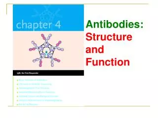
Antibodies: Structure and Function 2011
Antibodies: Structure and Function 2011. Peter Burrows/Scott Barnum 406 Shelby/BBRB 842 4-6529/4-4972 peterb@uab.edu sbarnum@uab.edu. Learning Objectives. To be able to understand: The structure of IgG Heavy and light chains Constant and variable regions

Antibodies: Structure and Function 2011
E N D
Presentation Transcript
Antibodies: Structure and Function 2011 Peter Burrows/Scott Barnum 406 Shelby/BBRB 842 4-6529/4-4972 peterb@uab.edu sbarnum@uab.edu
Learning Objectives • To be able to understand: • The structure of IgG • Heavy and light chains • Constant and variable regions • The concept of antibody classes and their biological functions • Polyclonal and monoclonal antibodies
You become infected with the flu virus IgM, IgA IgG, IgE
General Overview • Antibodies (Ab) are antigen (Ag)-binding proteins • Ab are present on the B-cell membrane AND are secreted by plasma cells • Membrane-bound Ab on a B cell confers specificity • Ag-specific proliferation of B cell clones depends on interaction of membrane Ab and Ag • Secreted Ab from plasma cells circulate in the blood and tissues or bathe mucosal secretions • These Ab serve as effectors of humoral immunity by neutralizing, eliminating, or preventing uptake of Ags
You become infected with the flu virus IgM, IgA IgG, IgE
Identification of the serum-protein fraction containing Abs (late 1930’s) • Tiselius and Kabat hyperimmunized rabbits, collected serum and then performed electrophoresis (separates proteins based on their overall charge). • Total serum (Blue) had four distinct peaks • (Black) Serum that was first incubated with the antigen to remove any antigen-binding proteins: significant reduction in g-globulin peak • The g-globulin fraction was identified as containing serum Abs and called immunoglobulins (Ig)
Elucidation of the basic structure of the immunoglobulin (IgG) molecule • Edelman and Porter used ultracentrifugation and found that the g-globulin fraction had a MW of ~150,000 • Papain digestion produced 2 identical fragments (MW 45,000) called Fab that retained Ag-binding and 1 Fc fragment (MW 50,000) • Pepsin digestion resulted in 1 Fab-like fragment (MW 100,00) called F(ab’)2 that retained Ag-binding • Mercaptoethanol reduction and alkylation (cleaves S-S bonds) revealed that the 150,000 molecule was composed of 2 identical 50,000 MW polypeptide chains (H-chains) and 2 identical 25,000 MW chains (L-chains) =
Amino Acid Sequencing Studies of Ig Heavy and Light chains - I • Light chain (L-chain): sequencing of many myeloma L-chains demonstrated that the N-terminal portions (100-110 aa) were variable (V region), whereas in the C-terminal portion there were only 2 types of sequences (Constant region) of k or l types • Heavy chain (H-chain) sequencing demonstrated that the N-terminal regions (100-110 aa) were variable among different H-chains (VH region), whereas the rest of the protein had 5 basic sequence patterns (m, g, a, d, e) and corresponded to 5 different H-chain constant regions called isotypes
Amino Acid Sequencing Studies of Ig Heavy and Light chains - II • The H-chain of an Ab defines the class (isotype) of that Ab: IgM (m), IgG (g), IgA (a), IgD (d), IgE (e) • Each isotype can have either korl L-chains • Each Ab molecule has 2 identical L-chains and 2 identical H-chains
Variable Region Domains of H- and L-chains • Amino acid sequences of V-light and V-heavy domains revealed variability is concentrated in several hypervariable regions (HV) or complementary-determining regions (CDRs) • Three HV regions are present in both H- and L-chains • HV regions form the Ag-binding site of Abs
Heavy Chain Lysozyme Antigenic Determinant Epitope Light Chain Antigen-antibody Interaction Three dimensional structure Fab of IgG Antigen
Hinge Region Component of Ig • IgG, IgD, and IgA H-chains contain an extended peptide sequence between CH1 and CH2 domains that is called the hinge region • Hinge region is rich in proline residues which gives IgG, IgD, and IgA Fab arms flexibility when bound to Ag • Extended polypeptide region makes Ig bearing hinge region more susceptible to proteolytic enzymes
Immunoglobulin Classes • 5 classes of Ig are distinguished by unique protein sequences in the H-chain constant region • H-chain constant region of each Ig confers class-specific structural and functional properties
Immunoglobulin G (IgG) • The most abundant Ig in serum (~80% of total serum Ig) • A monomer consisting of 2 g H-chains and 2 k or 2 l L-chains • 4 subclasses of IgG: IgG1, IgG2, IgG3, IgG4 (human) • The 4 subclasses of IgG are encoded by different germ-line constant heavy chain genes • The 4 subclasses differ by size of hinge region and number and position of interchain S-S bonds between H-chains • Amino acid differences between subclasses affect functional properties: IgG1, IgG3, and IgG4 cross placenta; IgG3 most effective activator of complement; IgG1 and IgG3 bind with high affinity to Fc receptors on phagocytic cells
Immunoglobulin M (IgM) Membrane-bound IgM Cµ3 • IgM - 5 to 10% of total serum Ig • IgM is a large pentamer in which 5 IgM monomers are held together by S-S bonds • IgM has 10 potential Ag-binding sites • Each pentamer of IgM contains Fc-linked polypeptide J-chain (joining chain) which is S-S linked • J-chain required for correct polymerization • IgM is the first Ab produced during a primary immune response • Monomeric IgM is the antigen receptor on B cells Cµ4 Secreted IgM
Immunoglobulin A (IgA) • IgA - 10-15% of serum Ig • Two subclasses IgA1 and IgA2 in humans • IgA is the predominant Ig class in mucosal secretions • Exists primarily in monomeric (serum) or dimeric/tetrameric (secretions) • IgA in secretions is referred to as secretory IgA (S-IgA) and is composed of IgA, J-chain, and a portion of Poly-Ig receptor • 5 to 10 g of S-IgA is secreted/day. In terms of production it is the highest. • Plasma cells that produce S-IgA preferentially migrate to sub-epithelial region of mucosal surfaces
Formation and transport of secretory IgA (S-IgA) • Polymeric-Ig receptor (pIgR) is expressed on basolateral surface of most mucosal epithelia tissue and glandular epithelia of mammary, lacrimal, and salivary glands • Dimeric IgA binds to the pIgR and is internalized • After transport of the pIgR-IgA complex to the lumenal surface, the receptor is cleaved and a portion (secreoty component/SC) remains bound to IgA resulting in formation of S-IgA • SC masks sites proteolytically sensitive sites within the hinge region which increase S-IgA stability in mucosal secretions • S-IgA serves as the main effector Ab in mucosal secretions
Immunoglobulin E (IgE) • Very low serum levels (0.3 mg/ml) • IgE mediates Type 1 hypersensitivity reactions (allergy, asthma, hay fever, hives, anaphylactic shock) • IgE binds Fc receptor of basophils and mast cells
Immunoglobulin E (IgE) • Very low serum levels (0.3 mg/ml) – IgE is pre-loaded onto mast cells • IgE mediates Type 1 hypersensitivity reactions (asthma, hay fever, hives, anaphylactic shock) • IgE binds Fce receptor of basophils and mast cells • Cross-linking of receptor bound IgE by Ag induces degranulation and release of histamine and other mediators • IgE may also participate in anti-parasitic defenses
Immunoglobulin D (IgD) Mature B cell IgM IgD • IgD levels in serum are low (~ 30 mg/ml) • IgD along with IgM are the major membrane-bound Ig • IgM and IgD are co-expressed on mature B cells • No known effector function for secreted IgD
Definition and generation of polyclonal and monoclonal antibodies • Most Ags possess multiple epitopes and induce activation, proliferation, and differentiation of many B-cell clones. The Abs produced are heterogeneous • The Ab response to an Ag which induces a mixture of Abs is referred to as a polyclonal Ab response. This Ab heterogeneity is good - increases immune protection • For research, diagnostic, and therapeutic purposes, it is often necessary to have an Ab to a single epitope of an Ag • The Ab response to a single epitope is referred to as a monoclonal Ab (mAb).
Definition and generation of polyclonal and monoclonal antibodies • Direct biochemical purification of a monoclonal Ab from a polyclonal Ab preparation is not feasible. • Köhler and Milstein devised a method to prepare mAbs by fusing a normal Ab-producing B cell with a plasmacytoma cell and yield a hybrid cell called a hybridoma • The hybridomas have immortal growth from the plasmacytoma and secreted the single type of Ab produced by the fused normal Ab-producing B cell.
Basic method to obtain monoclonal Ab • To select for fused cells: need myeloma cells that are HGPRT- and Ig-. These cells die in HAT media. Only fused cells derived from spleen B cells are HGPRT+ and Ig+. • After cell fusion, culture in HAT media for 7 to 10 days in which most cells are dead except the hybridomas. • Cells are subcultured at limiting dilution and screened for Ab of interest
Summary - I • The g-globulin fraction of serum contains antibodies, also referred to as immunoglobulins (Ig). • Ig are composed of two identical heavy (HC) and two identical light (LC) chains. The N-terminal portions (100-110 aa) are variable and called V regions, whereas the C-terminal portions are constant within a given LC type (k or l) or HC isotype (m, d, g, a, and e). • The variability of V regions is concentrated in three hypervariable regions (HV) or complementary-determining regions (CDRs). The HV or CDRs form the Ag-binding sites of Ab • The H-chain of an Ab determines the class (isotype) of that Ab: IgA, IgM, IgD, IgE, IgG. Each isotype can have eitherk or l LC.
Summary - II • IgG, IgD, and IgA H-chains contain extended peptide sequence between CH1 and CH2 domains and is called hinge region rich in proline residues which gives their Fab arms flexibility when bound to Ag. • The 5 classes of Ig are distinguished by unique aa sequences in the H-chain constant region and are encoded by different genes. • IgG is the most abundant Ig in serum (~80% of total serum Ig) and is a monomer consisting of 2 g H-chains and 2 k or 2 l L-chains. • IgM accounts for 5 to 10% of total serum Ig. Serum IgM is a pentamer in which 5 IgM monomers are held together by S-S and thus has potentially 10 Ag-binding sites. Generally the first Ab produced during primary immune response. • IgA is the predominant Ig class in mucosal secretions. Exists primarily in monomeric (serum) or dimeric/tetrameric (secretions) forms. In secretions it is referred to as secretory IgA, which contains dimeric or tetrameric IgA, J-chain, and a portion of Poly-Ig receptor. • Plasma cells that produce IgA preferentially migrate to sub-epithelial tissue.
Summary - III • Poly-Ig receptor (pIgR) is expressed on basolateral surface of most mucosal epithelia tissue and glandular epithelia of mammary, lacrimal, and salivary glands; Dimeric IgA binds to pIgR and is internalized by the cell. After transport of the receptor-IgA complex to the lumenal surface, a portion of receptor is cleaved and a portion (secretory component/SC) remains bound to IgA resulting in formation of S-IgA. • SC masks proteolytically sensitive sites within the hinge region which increase S-IgA stability in mucosals secretions. S-IgA serves as the main effector Ab in mucosal secretions. • IgE mediates hypersensitivity reactions (allergy, asthma, hay fever, hives, anaphylactic shock). It binds the Fce receptor of basophils and mast cells. Cross-linking of receptor bound IgE by Ag (allergen) induces degranulation and release of histamine. IgE may also participate in anti-parasitic defenses • A response that induces a mixture of Abs to one Ag is referred to as a polyclonal Ab response and is good for protection from pathogens. • Ab derived from a single B cell and directed to a single epitope on an Ag is referred to as a monoclonal Ab. These are good for diagnostics and therapeutics.