Download
1 / 36
360 likes | 509 Vues
Pathomor phology of kidney diseases. As.-proff. V.Voloshyn. According:prof. Bodnar Ya.Ya.; prof. Sorokina I.V.; Frank Netter. Main aim of lecture. To find out reasons and mechanisms of development of nephropathy. To expose – macro- and microscopic displays of nephropathy .

E N D
Pathomorphologyof kidney diseases As.-proff. V.Voloshyn According:prof. Bodnar Ya.Ya.; prof. Sorokina I.V.; Frank Netter
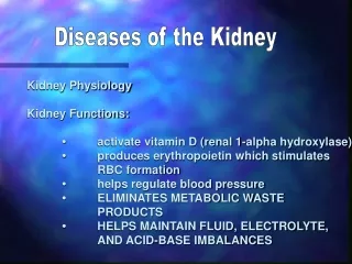
Main aim of lecture • To find out reasons and mechanisms of development of nephropathy. • To expose – macro- and microscopic displays of nephropathy. • To define the basic clinic-morphological displays of chronic kidney insufficiency. 2
Actuality • Illnesses of kidneys is group of illnesses which are the numeral and varied as in clinical so morphological displays and takes fifth line in morbidity of population after the traumatism, heart & vessels diseases, tumors and pulmonary pathologies. • Up to now the classification, diagnostics and treatments methods of nephropathy provocate the hot discussions between specialists of different type which diagnose and treat these illnesses 3
History of studies about pathomorphology of nephropathy • At the beginning of XVII century R. Brite offered the first classification of nephropathy • At the beginning of XX century clinicist Folgart and pathologist Far divided Brite’s illness on three groups: • - nephrites; • - nephroses; • - nephroscleroses. 4
Classification of nephropathy Structural-functional principle lies in the basis of modern clinic-morphologic classification of nephropathy. SELECT SUCH GROUPS OF NEPHROPATHY: • glomerulopathy; • tubulopathy; • tubulostromal nephropathy; • pyelonephritis; • nephrosclerosis • anomalies of development; • tumours. 5
Clinical syndromes at nephropathy • Urinary – is the recidive unpain hematuria, oliguria, proteinuria, leucocyturia, cylindruria; • Heart & vessels – is increase of arteriotony (especially diastolic), hypertrophy of the left ventricle of heart; • Nephrotic – is lipidemia, heavy proteinuria, warm white edemata of lower limbs. • Anaemic – toxic influence on marrow (firm (стійка) anaemia) 6
Introduction in practical work of nephrologists: - transskin kidney biopsy; - electronic microscopy; - іmmunomorphology The seat of pathomorphology in diagnostics of nephropathy 7
Glomerulopathy • Group of kidneys glomerules diseases which has congenital, primery-purchased and secondary-purchased origin. Their pathogeny consists in formation of immune complexes with their fixing on the basale membranes of glomerulis, or (in the case of autoimmune mechanism) formation of autoantibodies to the basale membrane. 9
Classification of glomerulopathy • Congenital glomerulopathy: • Syndrome of Alpoport • Congenital nephrotic syndrome • Inherited (спадковий) lardaceous? (amyloidosis) of kidneys • Initially-purchased glomerulopathy: • glomerulonephrytis with the minimum changing. • Postinfectial (poststreptococcic). • Subacute (semilunar). • Syndrome of Goodpascher. • Chronic glomerulonephrytis 11
Congenitalglomerulopathy • The syndrome of Alpoport is combination of lipoid tubulo- and interstitial nephropathy and glomerulopathy with the lowering of hearing and vision. • The inherited nephrotic syndrome is combination of minimum changing of podocytes, intracapillar productive glomerulonephrytis with polycystosis • Periodic illness – is combination of widespread lardaceous with polyserositis 12
Glomerulonephrytis with the minimum changing is characterized by practically complete absence of changes in preparation which are investigated by light microscope. An electronic microscopy displays out the changing of small appendices of podocytes 14
Postinfection (poststreptococcy) glomerulonephrytis The most frequent reason is В-hemolytic streptococcus of A group. • Glomerulis are megascopic, amemic, polymorphonuclear leucocytes are present in an interstitium. • An electronic microscopy displays out subephithelial deposits, proliferation and swelling of endotheliocytes 15
Subacute (quick progressic, malignant, semilunar glomerulonephrytis) “Large white kidney”, ”large pied (строкатa) kidney” ”large red kidney”. There are “crescent” (“halfmoons”) 16
Syndrome of Goodpascher • Quick progressicglomerulonephrytis which is combined with hemorrhagic-fibrinic pneumonia, and also with iron-deficient anemia. • Specific antibodies against the glicoprotein antigens of uncollogen portion of collogen IV are determined in a whey (сироватka) • In kidneys IgG and C3 are determined at immunofluorescence; At light microscopic – are “halfmoons”. 17
Chronic glomerulonephrytis • The independent disease is with the primary damage of glomerulis and secondary the pathological process extends into tubulis and stroma 18
Morphological forms of chronic glomerulonephrytis • Mesangioproliferativic • Membranous • Membranous-proliferativic or Mesangiocapillaric. • IgA is nephropathy (illness of Berje) 19
Mesangioproliferativic chronic glomerulonephrytis • Pathogeny is unknown • Proliferation of mesangiocytes is aspecific • Focal and segmental proliferation are simultaneous • The increasing of mesangial matrice At shick-reaction 20
Membranous chronic glomerulonephrytis Etiology • There is a idiopathic illness in 85 % of all patients. • Reasons of secondary membranous chronic glomerulonephritis are: • infections (syphilis, malaria, hepatitis B); • medicinal and chemical matters (penicillin, gold, mercury, heroin); • tumors (lymphadenomas, bronchogenic tumors); • sickle-cell anemia; • system lupus erythematosus. 21
Membranous chronic glomerulonephrytis • 1 stage - the subephithelial laying of deposits • 2 stages - the membranous and subephithelial laying of deposits • 3 stages - the intramembranous and mesangial laying of deposits • 4 stages - the diffuse laying of deposits with deformation of glomerular barrier 22
Mesangioproliferative chronic glomerulonephrytis • mesangial laying of deposits; • Ig G and C3 accumulates in mesangium more frequently. • focal proliferation of mesangiocytes • it is observed at primary IgA-nephropathy 23
IgA-nephropathy (illness of Berje) • most widespread reason of chronic kidney insufficiency • IgA is revealed in a whey • mesangial laying of deposits • IgA accumulates in mesangium • proliferation of mesangiocytes 24
Secondary acquisition glomerulonephrytes • Diabetic nephropathy • Amyloidosis of kidneys • Glomerulonephrytis at system lupus erytematosis 25
Secondary acquisition glomerulopathy(Diabetic nephropathy) 26 • Thickening of capillaries walls • Enlargement of volume of mesangium • Knot glome-rulosclerosis (changing of Kimmelsteel-Willson)
Secondary acquisition glomerulopathy(Amyloidosis of kidneys) • Greasy kidney • Accumulation of АА-amyloid in mesangium, basale membranes of vessels and tubulis • Positive reaction on an amyloid • Phasicness of motion: latent, proteinuric, nephrotic, nitrogenemic 27
Secondary acquisition glomerulopathy(System lupus erytematosus) • Laying of deposits in subepithelial and subendothelial layers • “ecraseurs” • formation of hematoxylin bodies 28
Secondary acquisition glomerulopathy(hemolytic-uremic syndrome) • Subendothelial laying of deposits • Thickening of capillaries walls • Thromboses • Infarcts of kidney cortex 29
Tubulopathy Purchased and inherited defeats of epithelium of nephron’s tubules, that is accompanied by violations of their concentric, reabsorb and secretory function. • Inherited tubulopathy - is predefined by the various forms of fermentopathies. • Purchased tubulopathy are: (acute kidney insufficiency, myelome of kidney, gouty kidney) 30
Acute kidney insufficiency 31 Necrosis of tubules is the morphological substratum of acute kidney insufficiency • Distinguish ischemic (shock) and toxic acute kidney insufficiency. • Reasons of toxic acute kidney insufficiency are the action: ---heavy metals (lead, mercury, bismuth, arsenic) of gold and uranium; ---organic solvents (chloroform, three-plus-onechlorous carbon); ---glycols (ethylen- and propilen-glycols); ---sulfanilamides, antibiotics (methacyclines, polimexinum); asteroid antiinflammation preparations; mercury diuretics
Tubuli-interstitial nephrite • Analgesic (analgin, aspirin in connection with a phenacetin): • Hypokaliumemial • Uratic (gouty) • Hypercalciumemial (metastatic calciphylaxis); • Radiation 32
Pyelonephritis Successive heterospecific bacterial ascending or descending inflammation of parenchima of kidney with predominant localization of pathological process in intermediate tissue. • Risk factors : short and wide urethra at women, stasis of urine of diverse etiology,urinary bladde-ureter reflux, age and hormonal status of patient • Women are ill mainly (1:5). 33