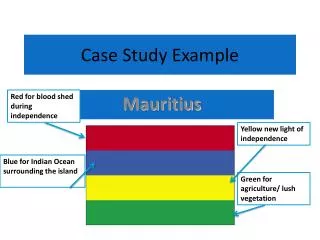
Example Case
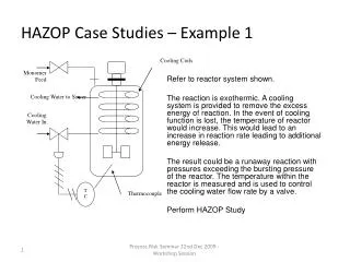
Transient Ischemic Attacks Rodney W. Smith, MD Clinical Assistant Professor Department of Emergency Medicine University of Michigan Ann Arbor, MI. Example Case. A 55 year old male presents to the emergency department with acute onset of Left arm weakness: Unable to lift left arm off of lap

Example Case
E N D
Presentation Transcript
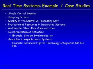
Transient Ischemic Attacks Rodney W. Smith, MDClinical Assistant ProfessorDepartment of Emergency MedicineUniversity of MichiganAnn Arbor, MI
Example Case • A 55 year old male presents to the emergency department with acute onset of • Left arm weakness: Unable to lift left arm off of lap • Symptoms improved on the way to the hospital
Example Case • PMHx: Hypertension • Takes enalapril • ROS: • No headache • No other neurologic symptoms • Social Hx: • Smokes 1 ppd
Example Case • Physical Exam • Overweight, in NAD • 160/90, 80, 14, 37.5C • Right carotid bruit • Heart with regular rate and rhythm; No murmur
Example Case • Neuro exam • Oriented to person, place, and time • Fluent speech • CN II-XII intact • Motor 4/5 strength in left upper extremity • Sensory subjective decrease in pinprick in left upper extremity compared to the right • DTR +2 except at left biceps +3 • Gait steady • Cerebellar intact finger to finger and finger to nose • No extensor plantar response.
Summary • Importance of distinguishing TIA from other causes of transient “spells” • Essential elements include a careful history, physical exam, and CT scan • ED treatment and disposition are directed toward prevention of subsequent stroke • Incidence of early stroke after TIA justifies hospital admission for further evaluation
Risk Factors/Epidemiology • 300,000 TIAs per year in US • 5-year stroke risk after TIA 29% • 43.5% in 2 years with >70% carotid stenosis treated medically • Many stroke patients have had TIA • 25% - 50% in large artery atherothrombotic strokes • 11% - 30% in cardioembolic strokes • 11% to 14% in lacunar strokes
Risk Factors/Epidemiology • Risk factors are the same as stroke • Increasing age • Sex • Family history / Race • Prior stroke / TIA • Hypertension • Diabetes • Heart disease • Carotid artery / Peripheral artery disease • Obesity • High cholesterol • Physical inactivity
ED Presentation • What is a TIA? • Acute loss of focal cerebral function • Symptoms last less than 24 hours • Due to inadequate blood supply • Thrombosis • Embolism
ED Presentation • Acute loss of focal cerebral function • Motor symptoms • Weakness or clumsiness on one side • Difficulty swallowing • Speech disturbances • Understanding or expressing spoken language • Reading or writing • Slurred speech • Calculations
ED Presentation • Acute loss of focal cerebral function • Sensory symptoms • Altered feeling on one side • Loss of vision on one side • Loss of vision in left or right visual field • Bilateral blindness • Double vision • Vertigo
ED Presentation • Non-focal Symptoms (Not TIA) • Generalized weakness or numbness • Faintness or syncope • Incontinence • Isolated symptoms (symptoms occurring alone) • Vertigo or loss of balance • Slurred speech or difficulty swallowing • Double vision
ED Presentation • Non-focal Symptoms (Not TIA) • Confusion • Disorientation • Impaired attention/concentration • Diminution of all mental activity • Distinguish from • Isolated language or visual-spatial perception problems (may be TIA) • Isolated memory problems (transient global amnesia)
ED Presentation • Acute loss of focal cerebral function • Abrupt onset • Symptoms occur in all affected areas at the same time • Symptoms resolve gradually • Symptoms are “negative”
ED Presentation • Symptoms last less than 24 hours • Most last less than one hour • Less than 10 percent > 6 hours • Amaurosis fugax up to five minutes
ED PresentationDifferential Diagnosis • Migraine with aura • Positive symptoms • Spread over minutes • Visual disturbances • Somatosensory or motor disturbance • Headache within 1 hour
ED PresentationDifferential Diagnosis • Aura without Headache • 98% Visual symptoms • 30% with other symptoms • 26% sensory • 16% aphasia • 6% dysarthria • 10% weakness • Mean age 48.7 (vs. 62.1) • Fewer cardiovascular risk factors
ED PresentationDifferential Diagnosis • Partial (focal) seizure • Positive sensory or motor symptoms • Spread quickly (60 seconds) • Negative symptoms afterward (Todd’s paresis) • Multiple attacks
ED PresentationDifferential Diagnosis • Transient global amnesia • Sudden disorder of memory • Antegrade and often retrograde • Recurrence 3% per year • Etiology unclear • Migraine • Epilepsy (7% within 1 year) • Unknown
ED PresentationDifferential Diagnosis • Transient global amnesia • No difference in vascular risk factors compared with general population • Fewer risk factors when compared with TIA patients • Prognosis significantly better than TIA
ED PresentationDifferential Diagnosis • Structural intracranial lesion • Tumor • Partial seizures • Vascular steal • Hemorrhage • Vessel compression by tumor
ED PresentationDifferential Diagnosis • Intracranial hemorrhage • ICH rare to confuse with TIA • Subdural hematoma • Headache • Fluctuation of symptoms • Mental status changes
ED PresentationDifferential Diagnosis • Multiple sclerosis • Usually subacute but can be acute • Optic neuritis • Limb ataxia • Age and risk factors • Signs more pronounced than symptoms
ED PresentationDifferential Diagnosis • Labyrinthine disorders • Central vs. Peripheral vertigo • Ménière's disease • Benign positional vertigo • Acute vestibular neuronitis
ED PresentationDifferential Diagnosis • Metabolic • Hypoglycemia • Hyponatremia • Hypercalcemia • Peripheral nerve lesions • Entrapments • Painful quality
ED PresentationDifferential Diagnosis • Patient evaluation by senior neurologists with interest in stroke • Agreement on 48 of 56 patients (85.7%) • 36 with TIA • 12 Not TIA • 8 of 56 disagreement • 4 of these, both listed firm diagnosis
ED Diagnosis and Evaluation • History • Characteristics of the attack • Associated symptoms • Risk factors • Vascular Disease • Cardiac Disease • Hematologic Disorders • Smoking • Prior TIA
ED Diagnosis and Evaluation • Physical Examination • Neurologic Exam • Carotid Bruits • Cardiac Exam • Peripheral Pulses
ED Diagnosis and Evaluation • EKG • CBC, Coags, and Chemistries • Chest Xray • Head CT without contrast • Expedite if early presentation
ED Diagnosis and Evaluation • Symptom vs. Disease • Significant carotid artery stenosis • Cardiac embolism • Admission vs. Discharge • Traditional approach • Trend toward outpatient evaluation
ED Diagnosis and Evaluation • Stroke Rate After TIA • Percent (95% CI)
ED Diagnosis and Evaluation • Stroke Rate After TIA • Johnston, et al. JAMA 284:2901, 2000. • Follow-up of 1707 ED patients diagnosed with TIA • Stroke rate at 90 days was 10.5% • Half of these occurred in the first 48 hours after ED presentation
Management • Goal: Prevention of Stroke • Expedited Evaluation • Carotid Artery Disease • Cardioembolism • Inpatient vs. Observation Unit vs. Outpatient • Antiplatelet Therapy • Risk Factor Modulation
ManagementED Disposition • Discharge • Further testing will not change treatment • Prior workup • Not a candidate for CEA or anticoagulation
ManagementED Disposition • Admission • Clear indication for anticoagulation • Severe deficit • Crescendo symptoms • Other indication for admission • Admission or observation unit evaluation • All others
ManagementDiagnosis of Carotid Stenosis • Carotid Duplex Ultrasound • Sensitivity of 94 - 100% for > 50% stenosis • May overdiagnose occlusion • Non-invasive
ManagementDiagnosis of Carotid Stenosis • Magnetic Resonance Angiography • Similar sensitivity to carotid ultrasound • Overestimates degree of stenosis • Gives information about vertebrobasilar system • Accuracy of 62% in detecting intracranial pathology • Cost and claustrophobia
ManagementDiagnosis of Carotid Stenosis • Cerebral Angiography • Gold standard for diagnosis • Invasive, with risk of stroke of up to 1% • For patients with positive ultrasound • For patients with occlusion on ultrasound • First test if intracranial pathology suspected
ManagementCardiogenic Embolism • Major risk factors: Anticoagulation Indicated • Atrial fibrillation • Mitral stenosis • Prosthetic cardiac valve • Recent MI • Thrombus in LV or LA appendage • Atrial myxoma • Infective endocarditis (No anticoagulation) • Dilated cardiomyopathy
ManagementCardiogenic Embolism • Minor risk factors: Best treatment unclear • Mitral valve prolapse • Mitral annular calcification • Patent foramen ovale • Atrial septal aneurysm • Calcific aortic stenosis • LV regional wall motion abnormality • Aortic arch atheromatous plaques • Spontaneous echocardiographic contrast
ManagementEchocardiogram • Yield < 3% in undifferentiated patients • Higher with risk factors • TEE preferred • Specific treatment of many abnormalities unknown
ManagementEchocardiogram • Indications • Age < 50 • Multiple TIAs in more than one arterial distribution • Clinical, ECG, or CXR evidence suggests cardiac embolization
Management TIA with Atrial Fibrillation • INR 2.5 (Range 2 to 3) • Aspirin if Warfarin contraindicated • Timing of onset of AC not proven in RCT • AC in other causes of cardioembolic stroke not proven in RCT EAFT Study Group, Lancet, 1993
ManagementAntiplatelet Therapy • Aspirin • Compared with placebo in patients with minor stroke/TIA • Relative risk of composite endpoint reduced by 13% to 17% • Dose of aspirin probably not important • Lower dose gives lower incidence of GI side effects.
Management • Ticlopidine • Small absolute risk reduction compared with ASA • Side effects preclude use in up to 5% • Serious adverse effects • Neurtropenia • Thrombotic thrombocytopenic purpura
Management • Clopidogrel • Similar to Ticlopidine in reducing composite endpoint • Reduction in risk of stroke alone less than with Ticlopidine • Similar side effect profile to ASA