Download
1 / 36
380 likes | 490 Vues
Integrative Physiology II: Fluid and Electrolyte Balance. 20. Mass Balance in the Body. Homeostasis requires that amounts gained must be equal to that lost. Ion concentration- need proper amounts of Na+, Cl-, K+, and Ca2+:

E N D
Mass Balance in the Body • Homeostasis requires that amounts gained must be equal to that lost. • Ion concentration- need proper amounts of Na+, Cl-, K+, and Ca2+: • nervous, cardiac& muscle function-imbalances cause problems with membranes of cells that are excitable. • Primarily replaced with thirst & appetite and excreted in urine, sweat, & feces • pH balance- cells functions within a pH range that is maintained by H+, CO2, & HCO3– • Fluid- water levels need to be maintained, ingestion and urine formation have largest impact.
Key factors for Homeostasis • Na+ & H2O= affect ECF • Osmolarity: amounts of solutes dissolved in solution, concentration and permeability influence direction of osmosis which changes the size of cells
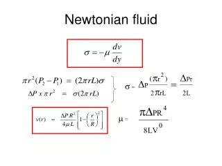
Osmosis and Osmotic Pressure Osmolarity describes the number of particles of solution in a quantity of osmoles OsM per liter Osmolarity is influence by fluid, ion, & protein levels. Compensation occurs via renal, behavioral, repiratory, and CV responses Figure 5-29
Water Balance in the Body Water makes up 50-60% of total body weight. Main entry way is through food, most lost in urine unless there is excessive sweating or diarrhea Homeostasis maintains water balance unless there is pathology or an abnormal ingestion of water. Men have more water than women Figure 20-2
Water Balance A model of the role of the kidneys in water balance Kidneys cannot add water, only preserve it or get rid of excess amounts. Renal filtration will stop if there is a major loss causing extremely low blood pressure and blood volume Figure 20-3
Fluid and Electrolyte Homeostasis The body’s integrated response to changes in blood volume and blood pressure incorporate many systems Decreased blood volume will result in mechanisms that increase blood pressure and volume, and reduce water loss Figure 20-1a
Fluid and Electrolyte Homeostasis Increased blood volume results in the excretion of salt and water which eventually reduces blood pressure and ECF/ICF volumes. Figure 20-1b
Urine Concentration Osmolarity changes as filtrate flows through the nephron. Reabsorption is controlled by kidney tissue concentrations as water permeability and diffusion of solutes changes as needed. Water and sodium reabsorption alter urine concentration. Diuresis is the removal of excess water. Figure 20-4
Water Reabsorption Vasopression or antidiuretic hormone causes a graded effect of forming water pores on collecting duct cells. Thus permeability is increased and more water is retained making urine more concentrated. Figure 20-5a
Water Reabsorption If vasopressin is absent water will not move out through water pores (aquaporins)and the urine will be dilute. Figure 20-5b
Water Reabsorption Cross-section of kidney tubule Medullary interstitial fluid Collecting duct lumen Vasa recta Collecting duct cell 600 mOsM Filtrate 300 mOsm 600 mOsM 700 mOsM Second messenger signal 2 1 Vasopressin cAMP Vasopressin receptor Vasopressin binds to mem- brane receptor. Receptor activates cAMP second messenger system. 1 2 The mechanism of vasopressin action on tubular cells of the nephron Figure 20-6, steps 1–2
Water Reabsorption Cross-section of kidney tubule Medullary interstitial fluid Collecting duct lumen Vasa recta Collecting duct cell 600 mOsM Filtrate 300 mOsm 600 mOsM 700 mOsM Storage vesicles Second messenger signal 2 Exocytosis of vesicles 1 Vasopressin Aquaporin-2 water pores cAMP 3 Vasopressin receptor Cell inserts AQP2 water pores into apical membrane. Vasopressin binds to mem- brane receptor. Receptor activates cAMP second messenger system. 1 2 3 Apical membrane water permeability increases exponentially with water pores are added Figure 20-6, steps 1–3
Water Reabsorption Cross-section of kidney tubule Medullary interstitial fluid Collecting duct lumen Vasa recta Collecting duct cell 600 mOsM Filtrate 300 mOsm H2O 600 mOsM H2O H2O H2O 700 mOsM 4 Storage vesicles Second messenger signal 2 Exocytosis of vesicles 1 Vasopressin Aquaporin-2 water pores cAMP 3 Vasopressin receptor Vasopressin binds to mem- brane receptor. Receptor activates cAMP second messenger system. Cell inserts AQP2 water pores into apical membrane. Water is absorbed by osmosis into the blood. 4 1 2 3 Vassopressin is also called antidiuretic hormone- it causes reabsortion of water (in turn increasing urine concentration and decreasing volume). Figure 20-6, steps 1–4
Factors Affecting Vasopressin Release Three stimuli control vasopressin but the most potent is blood osmolarity above 280mOsM. The higher the osmolarity, the more vasopressin released by posterior pituitary. Osmoreceptors also trigger thrist centers in hypothalamus Figure 20-7
Water Balance Countercurrent exchange in the medulla of the kidney. Descending limb is permeable to water while the ascendig limb is permeable to ions. 25% of all Na+ and K+ reabsorption happens in ascending limb; resulting in dilute urine. Water amounts can be changed again at distal tubule and collecting duct. Figure 20-10
Fluid and Electrolyte Balance • Vasa recta removes water-blood runs in opposite direction as filtrate, water moves in according to the concentration gradient • Close anatomical association of the loop of Henle and the vasa recta-this allows for water to move out of the tubule and into the blood without dilution the interstitial fluid in the medulla. • Urea increase the osmolarity of the medullary interstitium-transporter proteins move urea into the medulla to increase osmolarity of the interstitial fluid creating a gradient to move water out without affecting the movement of other ions (Na+ & K+)
Sodium Balance Homeostatic responses to salt ingestion show the integrated effects on sodium, water, and blood pressure. Without salt appetite [salt] would increase and tissue cells would shrink. Thus vasopressin and thirst is activated. . Figure 20-12
Sodium Balance Aldosterone combines with a cytoplasmic receptor. 1 Interstitial fluid P cell of distal nephron Blood 2 Hormone-receptor complex initiates transcription in the nucleus. Lumen of distal tubule Transcription Aldosterone 2 1 mRNA Aldosterone receptor • Aldosterone action in principle cells- targets cells of the distal convoluted tubule and collecting duct. Figure 20-13, steps 1–2
Sodium Balance Aldosterone combines with a cytoplasmic receptor. 1 Interstitial fluid P cell of distal nephron Blood 2 Hormone-receptor complex initiates transcription in the nucleus. Lumen of distal tubule Transcription Aldosterone 2 1 mRNA Translation and protein synthesis 3 Aldosterone receptor 3 New protein channels and pumps are made. New channels New pumps ATP 4 Proteins modulate existing channels and pumps. Aldosterone- induced proteins modify existing proteins. 4 ATP Existing channels are called the leak proteins that allow for a rapid movement of ions Figure 20-13, steps 1–4
Sodium Balance Aldosterone combines with a cytoplasmic receptor. 1 Interstitial fluid P cell of distal nephron Blood 2 Hormone-receptor complex initiates transcription in the nucleus. Lumen of distal tubule Transcription Aldosterone 2 1 mRNA Translation and protein synthesis 3 Aldosterone receptor 3 New protein channels and pumps are made. New channels New pumps ATP 4 Proteins modulate existing channels and pumps. Aldosterone- induced proteins modify existing proteins. 4 K+ secreted K+ K+ K+ 5 ATP Na+ reabsorbed Na+ Na+ Result is increased Na+ reabsorption and K+ secretion. 5 Na+ Aldosterone causes K+ secretion and sodium reabsorption. A secondary effect is that water follows sodium. Figure 20-13, steps 1–5
Sodium Balance Decreased blood pressure stimulates renin secretion. Granular cells can be activated to release renin by three factors: drop in blood pressure, a signal from the kidneys, or increased sympathetic activity. Figure 20-15
Sodium Balance The renin-angiotensin-aldosterone pathway- (RAAS). Renin is an enzyme that assist in ANG II formation. ANGII activates several mechanisms that ultimately increase blood pressure and volume Figure 20-14
Sodium Balance Action of natriuretic peptides-cause sodium loss through urine (natriuresis) and act as RAAS antagonist. They are released when myocardial cells stretch too much or during heart failure Figure 20-16
Potassium Balance • Regulatory mechanisms keep plasma potassium in narrow range (3.5-5meq/L) • Aldosterone is released in response to excess levels, it increases permeability at distal nephron so K is moved into the urine while sodium is reabsorbed • Hypokalemia (K+ levels below 3) • In ECF levels are low, K+ leaves the cell, and resting membrane potential is more negative (hyperpolaized)= stronger stimulus • Muscle weakness and failure of respiratory muscles and the heart due to hyperpolarized neurons. • Hyperkalemia (K+ levels above 6) • In ECF levels are high, more K+ enters the cell, thus depolarizing it but then less able to repolarize thus LESS excitable • Can lead to cardiac arrhythmias • K+ irregularities include kidney disease, diarrhea, and diuretics
Behavioral Mechanisms Drinking water and eating salt is the only way the body obtains these substances, therefore individuals who cannot do this must be assisted. • Drinking replaces fluid loss – when body osmolarity raises above 280mOsM hypothalmic osomreceptors trigger thrist. Oropharynx receptors are stimulated by cold drink and signal thirst quench • Low sodium stimulates salt appetite – the hypothalamus also has centers for salt appetite which trigger a response when osmolarity is low. • Avoidance behaviors help prevent dehydration • Desert animals avoid the heat
Disturbances in Volume and Osmolarity In each situation compensantion mechanism aim to bring conditions to normal, in some cases there is incomplete compenstation. Notice how most imbalances are due to what is ingested or loss in excess amounts Figure 20-17
Severe Dehydration Compensation • Condition: low ECF volume, low blood pressure, high osmolarity • Compensation Mechanisms • Cardiovascular Responses- increase cardiac output & vasoconstriction to increase blood pressure. Vasoconstriction reduces GFR activating granular cells to release renin • Angiotensin II- produce after renin release that activates RAAS pathway to trigger thirst, vasopressin release, and vasoconstrion. (aldosterone is not release as it would increase osmolarity) • Vassopressin- increase water reabsorption to reduce loss in urine • Thrist/ IV-replacement of loss fluids and lowering of osmolarity
Acid-Base Balance • Normal plasma pH is 7.38–7.42-also resembles the pH inside cells, its optimum for proper protein & enzyme function • H+ concentration is closely regulated-slight pH changes indicate a 10-fold increase/decrease in [H+] which can have damaging effects on protein structure & function. • Abnormal pH affects the nervous system- H+ imbalances cause K+ imbalances because transporter protein in kidneys moves H+ and K+ in antiport fashion • Acidosis: neurons become less excitable and CNS depressionpatients can fall into a coma or have respiratory failure • Alkalosis: hyperexcitable- numbness, tingling, muscle twitches, severe cases lead to paralysis of respiratory muscles • pH disturbances- induced by an imbalance of H+ input/output • Compensation by buffers, ventilation, or renal regulation • Greatest source is CO2 level changes induced by metabolic or respiratory factors
Acid-Base Balance Hydrogen balance in the body is quickly compensated by ventilation and slowly compensated by renal regulation. Figure 20-19
Acid and Base Input Acid • Organic acids – acids produced by the body during metabolism or ingested molecules that realease H+ (acidic fruits, amino acids, fatty acids) • Under extraordinary conditions – produce more H+ than the body can normally get rid off and cause pathological effects • Metabolic organic acid production can increaseKetoacids – strong acids produced when fats & proteins are metabolized • Diabetes – metabolism disorder causes ketoacid formation • Accumulation of CO2- can occur as a result of anaerobic respiration, increased metabolism, or decreased ventilation • Acid production - CO2combines with water rapidly to make an acid and drop pH. Respiratory system gets rid of 75% of a Base • Few dietary sources of bases- conditions of alkalosis rarely encountered
pH Disturbances The reflex pathway for respiratory compensation of metabolic acidosis responds to shifts in H+ & CO2 based on the law of mass action. CO2 + H2O == H2CO3 == H+ HCO3 . Figure 20-20
pH Homeostasis • Buffers moderate changes in pH- cannot prevent changes, they absorb/release H+ • Cellular proteins, phosphate ions, and hemoglobin- serve as intracellular buffers • Ventilation • Rapid- quickly gets rid of CO2 by increasing breathing rate • 75% of disturbances- are cleared by ventilation thanks to the central and peripheral chemoreceptors that sense changes in [H+] • Renal regulation- uses ammonia and phosphate buffers in addition to • Directly excreting or reabsorbing H+ • Indirectly by change in the rate at which HCO3– buffer is reabsorbed or excreted
pH Disturbances Overview of renal compensation for acidosis. The nephron takes care of the 25% of compensation the lungs can’t handle. They excrete H+ by trapping it in ammonia and phosphate ions. They also make HCO3 Figure 20-21
Intercalated Cells Role of intercalated cells in acidosis and alkalosis – these are located in between principal cells of the distal tubule and have high amounts of carbonic anhydrase. Movement occurs via H+-ATPase and H+-K+-ATPase Figure 20-23
Acid-Base Balance Respiratory system increases CO2 during hypoventilation and decreases it during hyperventilation. When metabolism causes a disturbance respiratory keeps CO2 levels normal but pH changes because buffer levels drop. Mass balance shifts equation to left or right CO2 + H2O == H2CO3 == H+ HCO3 .