### Phylogenetic Analysis and Subcellular Localization of TaLTP Proteins in Wheat and Rice ###
This study presents a phylogenetic tree of TaLTPIb.1, TaLTPIb.5, and TaLTPId.1, utilizing putative 9 KDansLTP genes from NCBI for pairwise alignment through ClustalW. The eight-cysteine motif facilitated phylogenetic tree construction via MEGA 5.0, employing the Maximum Likelihood algorithm with 1,000 bootstrap replicates. Additionally, the effects of various hormone treatments (MeJA, SA, ABA, IAA) on wheat seedling phenotypes and the expression levels of TaLTPs were analyzed using RT-PCR. Subcellular localization studies were performed using TaLTPIb.1:EGFP fusion proteins in onion epidermal cells. ###

### Phylogenetic Analysis and Subcellular Localization of TaLTP Proteins in Wheat and Rice ###
E N D
Presentation Transcript
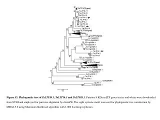
Figure S1. Phylogenetic tree of TaLTPIb.1, TaLTPIb.5 and TaLTPId.1. Putative 9 KDansLTP genes in rice and wheat were downloaded from NCBI and employed for pairwise alignment by clustalW. The eight cysteine motif was used for phylogenetic tree constructionby MEGA 5.0 using Maximum likelihood algorithm with 1,000 bootstrap replicates.
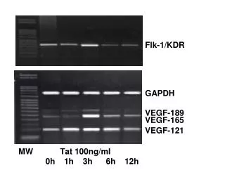
A B Control MeJA SA ABA IAA 0h6h12h24h TaLTPb.1 TaLTPb.5 0h TaLTPd.1 Ta18srRNA C 6h D 0h20min40min1h3h9h24h48h72h TaLTPb.1 TaLTPb.5 12h TaLTPd.1 Ta18srRNA E 24h • Figure S2RT-PCR conditions of TaLTPIb.1, TaLTPIb.5 and TaLTPId.1. (A) Phenotypes of wheat seedlings under MeJA (100 μM), SA (2 mM), ABA (100 μM), IAA (100 μM) treatments; (B) Semi-quantitative RT-PCR of TaLTPs under control conditions in hormone treatments (C) Real-time PCR of TaLTPs under control conditions in hormone treatments; (D) Semi-quantitative RT-PCR of TaLTPs under control conditions in abiotic stress treatments; (E) Real-time PCR of TaLTPs under control conditions in abiotic stress treatments.
Merge EGFP Bright Field A2 A3 A1 TaLTPIb.1:EGFP pm cw B2 B3 B1 35S:EGFP nu • Figure S3Subcellular localization of TaLTP:EGFP fusion proteins in onion epidermal cells. Onion epidermal cells transiently transformed with either the TaLTIb.1:EGFP (A) or EGFP control (B) were incubated in 0.8 M mannitol to induce plasmolysis and then imaged by confocal microscopy. pm, plasma membrane; cw, cell wall; nu, nuclear