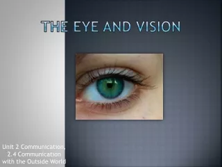

Eye and Vision
E N D
Presentation Transcript
Eye and Vision Exercise 26 BI 232
External Features • Notice the pupil which is surrounded by the colored iris. • The sclera is the white of the eye which is covered by a membrane called the conjunctiva • Eyelids come together at lateral and medial commissures. • The lacrimalcaruncle is found in the medial commissure.
Lacrimal Apparatus • Consists of the lacrimal gland and its accessory structures. • Produces water, alkaline tears. • Contain antibacterial enzyme called lysozyme for protection from bacterial infections
Extrinsic Eye Muscles • Superior oblique: primarily rotates the top of the eye toward the nose and secondarily moves the eye downward • Innervated by CNIV (trochlear) • Trochlea: Ligament sling • Superior rectus: primarily moves the eye upward and secondarily rotates the top of the eye toward the nose • Innervated by CNIII (oculomotor) • Lateral rectus: moves the eye away from the nose • Innervated by CNVI (abducens)
Extrinsic Eye Muscles • Medial rectus: moves the eye toward the nose • Innervated by CNIII (occulomotor) • Inferior oblique: primarily rotates the top of the eye away from the nose and secondarily moves the eye upward (also CNIII) • Inferior rectus: primarily moves the eye downward and secondarily rotates the top of the eye away from the nose (also CNIII)
Ear Nose
Ciliary body A • Ciliary processes • Ciliary epithelium • Secretes aqueous humor • Ciliary muscle - (intrinsic eye muscle) • Suspensory ligament of the lens A= anterior chamber P= posterior chamber P 3 1 2
Intrinsic Eye Muscles of the Iris • Pupils constrict (Parasympathetic) • Close vision and bright light • Pupils dilate (Sympathetic) • Distant vision and dim light
Neural pathway for vision • After optic nerves exit the eyeballs they meet at the optic chiasm. • Fibers from medial half of retina cross over to the opposite side. • Optic tracts project to the lateral geniculate bodies in thalamus • Some fibers are relayed to superior colliculi
Eye Dissection • Do eye dissection • Be able to ID structures for quizzes and practical
Determination of the Near Point • The minimum distance an object can comfortably be held in focus • Changes as we age • Hold a paper with fine print vertically at arm’s length in front of you. • Close one eye and slowly move the paper closer until either you see two objects or it becomes blurry. • Have partner measure distance
Distribution of Rods and Cones • Slowly move a small colored object, without letting your lab partner see it, from the back of your lab partner’s head, around the side toward the front. • With lab partner still looking straight ahead, not the angle when they are able to see the object.(rods) • Continue to move object forward slowly until color recognition occurs
Binocular Visual Field • Close right eye and look straight ahead with the left • Lab partner moves an object from behind your head from the left until you can see object. • Measure angle with protractor from tip of nose • Continue moving object until it disappears. • Repeat the exercise with the other eye • Determine the total visual field by adding the sums of above.
Visual Acuity • Snellen eye chart used to test visual acuity • 20/20 normal • 20/15 you can see at 20 feet what people with normal vision see at 15 feet. • 20/30 you see at 20 feet what people with normal vision see at 30 feet.
Astigmatism • The degree of curvature in the cornea or lens varies from one axis to another (is uneven or wavy) • This causes light to focus on more than one area of the retina creating a blurry image.
Ophthalmoscope • Used for diagnosis of variances in the eyeball but also as a potential indicator of diseases, such as diabetes mellitus. • Sit facing your lab partner • Have scope set at zero • Move in close to examine the eye of your partner • Look into the pupil
Refraction • Light is bent when it passes from one medium to another medium with a different density • Light passes through these before it hits the retina: • Cornea • Aqueous humor • Lens • Vitreous humor
Focal Point & Focal Distance • Focal Point: The specific point of intersection on the retina. • Focal distance: The distance between the center of the lens and its focal point. Determined by two factors: • Distance from the object to the lens • Shape of the lens
Focal Distance • Distance from the object to the lens: the closer an object is, the greater the focal distance • Shape of the lens: the rounder the lens, the more refraction occurs, so it has a shorter focal distance
Accommodation • Accommodation is an alteration in the curvature of the lens of the eye to focus an image on the retina • Near objects: Lens becomes rounder • Distance objects: Lens becomes flatter
Accommodation • Emmetropia is normal vision. • The image will be focused on the retina’s surface
Accommodation Problems • Myopia: Nearsighted • The eyeball is too deep or the curvature of the lens is too great • The focal point is in front of the retina, so distance objects are blurry • Corrected with a diverging lens
Accommodation Problems • Hyperopia: Farsighted • The eyeball is too shallow or the curvature of the lens is too flat • The focal point is behind of the retina, so near objects are blurry • Corrected with a converging lens
Color Blindness • Cones are responsible for color vision. • 3 types of cones each able to absorb light at specific wavelengths. • Lack of one or more types of cone can cause color blindness • Use Ishihara color plates to test
Afterimages • Photosensitive pigment of the rods is rhodopsin, a light-sensitive retinal and the protein opsin. • When light strikes the retina, rhodopsin splits into its two component parts and becomes pale (bleaching) • You can test the time for separation and reassembly of the photopigments by staring at a contrasting image on a card • Stare about 10-20 seconds and then shuts your eyes. Have your partner record time. You should see colored image against dark background (positive afterimage) After a few moments you should see the reverse of the original image (negative afterimage)
Blind Spot • The optic disc is a region where the nerve fibers exit the back of the eye and form the optic nerve. • This region is devoid of photoreceptors and is called the blind spot. • Follow the instructions from your instructor or book to find your blind spot
The End • Spend the rest of the lab locating structures on the eye models • Make sure that you understand the tests we did in class because you will be asked about them on quizzes and practicals