Download
1 / 24
240 likes | 370 Vues
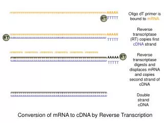
Involvement of Histone H1.2 in Apoptosis Induced by DNA Double-Strand Breaks. Konishi et al., Cell, Vol 114, 673-688, 2003. RCC1 ( inxs/Bj1 ). Regulator of Chromatin Condensation-1:

E N D
Involvement of Histone H1.2 in Apoptosis Induced by DNA Double-Strand Breaks Konishi et al., Cell, Vol 114, 673-688, 2003.
RCC1 (inxs/Bj1) Regulator of Chromatin Condensation-1: • Regulates chromatin condensation prior to entry into mitosis (normally inhibits condensation until replication is complete; in mutants, get premature chromatin condensation (PCC) before the completion of DNA replication) • Ran Guanine Nucleotide Exchange Factor (Ran GEF) required for Nuclear import (NLS proteins) and export (?) • Mammalian RCC1 binds histones H2A and H2B (Ref: ____) • Drosophila dRCC1 “associated with” nucleosomes (Frasch, 1991)
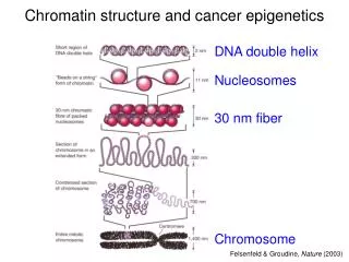
Major Histone Proteins G.M. Cooper, 2000 Histone H1: 8 isotypes in mice and humans H1.1-H1.5 are ubiquitously expressed in all cell types
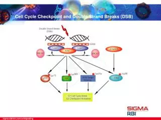
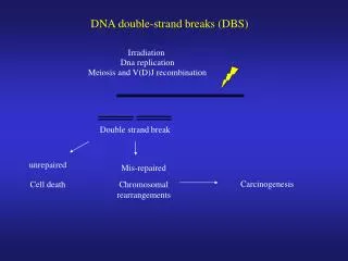
DNA damage-induced apoptosis • In response to DNA damage, p53 activates transcription of two cytoplasmic pro-apoptotic BH3-only proteins (Noxa and Puma) • The pro-apoptotic multidomain Bax is upregulated transcriptionally, the protein undergoes a conformational change, and is translocated to the mitochondrial membrane • P53, p53AIP1, TR3 (nuclear orphan receptor) and LKB1 translocate to the mitochondria • Translocation to the mitochondria induces mitochondrial membrane permeabilization which results in the release of apoptogenic factors (e.g. cytochrome C, AIF) into the cytoplasm that activate downstream • destruction programs
Mitochondria in Apoptosis SIGMA-ALDRICH Bax (pro-apoptotic) ? Smac/DIABLO Omi Endonuclease G 1. DNA Damage
Question:What are the mechanisms of signal transmission from the nucleus to the mitochondria during DNA damage-induced apoptosis? Approach: Search for a cytochrome C-releasing factor that appears in the cytoplasm after X-ray irradiation of the thymus.
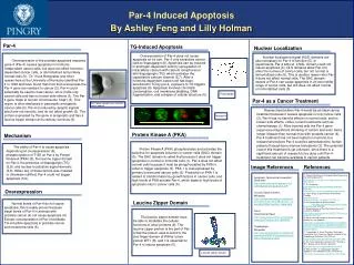
Histone H1.2 induces cytoC release from isolated mitochondria
Histone H1.2 Cyto-C release is Bak-dependent Oligomeric Bak (active) Monomeric Bak
Histone H1.2 accumulates in the cytoplasm (inxs – try in cultured cells and/or salivary gland?)
Histone H1.2 is released from the nucleus Leptomycin B inhibits apoptosis of irradiated cells; i.e. Nuclear Export is required for apoptosis (inxs – both import and export?) MCF7 cells: Functional p53 (irradiated) Q: Is histone H1 upregulated during X-ray induced apoptosis?
Reduced Histone H1.2 expression inhibits X-ray-induced apoptosis
H1.2-Deficient mice are resistant to apoptosis induced by X-ray & etoposide +/+ -/-
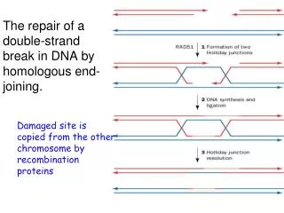
Discussion • DNA double-strand breaks, but probably not UV damage, trigger a megabase range of chromatin remodeling which leads to release of a significant amount of H1 from the chromatin • The amount of histone H1.2 released from the chromatin might be used for monitoring the extent of DNA damage • The release of nuclear histone H1 and its accumulation in the cytoplasm suggest that nuclear import/export machinery must be affected by X-ray irradiation (look at role of inxs in X-ray induced apoptosis also??) • How does H1.2 induce cytochrome C release? • The major difference between H1.2 and the other H1s is at the C-terminal region: KXXKP exists in all H1s except H1.2 (negative regulatory sequence?)
Caspase-dependent deubiquitination of monoubiquitinated nucleosomal histone H2A induced by diverse apoptogenic stimuli Mimnaugh et al., CDD, 2001.
Programmed Cell Death SIGMA-ALDRICH
Programmed Cell Death Programmed cell death (PCD), or apoptosis, can be triggered by a wide range of stimuli, including cell surface receptors like Fas and FasL. It constitutes a system for the removal of unnecessary, aged, or damaged cells that is regulated by the interplay of proapoptotic and antiapoptotic proteins of the Bcl-2 family. The proapoptotic proteins Bax, Bad, Bid, Bik, and Bim contain an -helical BH3 death domain that fits the hydrophobic BH3 binding pocket on the antiapoptotic proteins Bcl-2 and Bcl-XL, forming heterodimers that block the survival-promoting activity of Bcl-2 and Bcl-XL. Thus, the relative abundance of proapoptotic and antiapoptotic proteins determines the susceptibility of the cell to programmed death. The proapoptotic proteins act at the surface of the mitochondrial membrane to decrease the mitochondrial trans-membrane potential and promote leakage of cytochrome c. In the presence of dATP cytochrome c complexes with and activates Apaf-1. Activated Apaf-1 binds to downstream caspases, such as procaspase-9, and processes them into proteolytically active forms. This begins a caspase cascade resulting in apoptosis. References Kelekar, A., and Thompson, C.B., Bcl-2-family proteins: the role of the BH3 domain in apoptosis. Trends Cell. Biol., 8, 324-330 (1998). Priault, M., et al., Investigation of bax-induced release of cytochrome c from yeast mitochondria: permeability of mitochondrial membranes, role of VDAC and ATP requirement. Eur. J. Biochem., 260, 684-691 (1999). McDonnell, J.M., et al., Solution structure of the proapoptotic molecule BID: a structural basis for apoptotic agonists and antagonists. Cell, 96, 625-634 (1999).
Mitochondria in Apoptosis Increases in cytosolic Ca2+ levels due to activation of ion channel-linked receptors, such as that for the excitatory amino acid neurotransmitter glutamic acid, can induce permeability transition (PT) of the mitochondrial membrane. PT constitutes the first rate-limiting event of the common pathway of apoptosis. Upon PT, apoptogenic factors leak into the cytoplasm from the mitochondrial intermembrane space. Two such factors, cytochrome c and apoptosis inducing factor (AIF), begin a cascade of proteolytic activity that ultimately leads to nuclear damage (DNA fragmentation, DNA mutations) and cell death. Cytochrome c, a key protein in electron transport, appears to act by forming a multimeric complex with Apaf-1, a protease, which in turn activates procaspase 9, and begins a cascade of activation of downstream caspases. Smac/Diablo is released from the mitochondria and inhibits IAP (inhibitor of apoptosis) from interacting with caspase 9 leading to apoptosis. Bcl-2 and Bcl-X can prevent pore formation and block the release of cytochrome c from the mitochondria and prevent activation of the caspase cascade and apoptosis. PT is also related to the mitochondrial generation of reactive oxygen species which plays a role in the degradation phase of apoptosis (i.e. plasma membrane alterations). References Budihardjo, I., et al., Biochemical pathways of caspase activation during apoptosis. Annu. Rev. Cell Dev. Biol., 15, 269-290 (1999). Susin, S.A., et al., Molecular characterization of mitochondrial apoptosis-inducing factor. Nature, 397, 441-446 (1999). Cai, J., et al., Mitochondrial control of apoptosis: the role of cytochrome c. Biochim. Biophys. Acta, 1366, 139-149 (1998). Lee, H., and Wei, Y., Mitochondrial role in life and death of the cell. J. Biomed. Sci., 7, 2-15 (2000).