Download
1 / 27
270 likes | 413 Vues
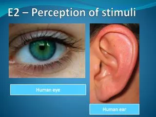
Perception of stimuli. Option E.2. Assessment statements. E.2.1 Outline the diversity of stimuli that can be detected by human sensory receptors, including mechanoreceptors, chemoreceptors, thermoreceptors and photoreceptors. E.2.2 Label a diagram of the structure of the human eye.

E N D
Perception of stimuli Option E.2
Assessment statements • E.2.1 Outline the diversity of stimuli that can be detected by human sensory receptors, including mechanoreceptors, chemoreceptors, thermoreceptors and photoreceptors. • E.2.2 Label a diagram of the structure of the human eye. • E.2.3 Annotate a diagram of the retina to show the cell types and the direction in which light moves. • E.2.4 Compare rod and cone cells. • E.2.5 Explain the processing of visual stimuli, including edge enhancement and contralateral processing. • E.2.6 Label a diagram of the ear. • E.2.7 Explain how sound is perceived by the ear, including the roles of the eardrum, bones of the middle ear, oval and round windows, and the hair cells of the cochlea.
Sensory receptors and diversity of stimuli • We have learned to link certain tastes, sights and sounds with emotions • Sensory cells send messages to certain parts of the brain that control emotion and memory • Nerve impulses arriving at the brain results in sensation
Mechanoreceptors • Stimulated by mechanical force or some type of pressure • Sense of touch is due to pressure receptors • Pressure receptors are also found in: • Arteries (detect change in blood pressure) • Lungs (stretch receptors respond to the degree of lung inflation) • Arms and legs (proprioceptors which tell us position and help maintain posture) • Inner ear (pressure receptors sensitive to waves of fluid moving over them)
Chemoreceptors • Respond to chemical substances • Allows us to taste and smell • Found in blood vessels and monitor pH • Pain receptors respond to chemicals released by damaged tissues
Thermoreceptors • Respond to change in temperature
Photoreceptors • Respond to light energy • Found in our eyes • Rod cells respond to dim light resulting in black and white vision • Cone cells respond to bright light and give us color vision
TOK • To what extent are we dependent on technology for our knowledge of biology? • http://physics.ucsd.edu/neurophysics/publications/New_Scientist.pdf
Structure of the human eye Posterior chamber pupil fovea Suspensory ligament Ciliary muscle
Cow eye dissection • http://www.exploratorium.edu/learning_studio/cow_eye/
The retina • Vision begins when light enters the eye and is focused on the photoreceptor cells of the retina • Both rods and cones synapse with their own bipolar neurons • Each bipolar neuron synapses with a ganglion cell • Axons of the ganglion cell make up the optic nerve which carries the message of vision to the brain
Annotation of retina diagram • Rods are photoreceptor cells which are sensitive to light and function well in dim light; synapse with a bipolar neuron • Cones are photoreceptor cells which are activated by bright light; synapse with a bipolar neuron • Bipolar neurons are cells in the retina which carry impulses from a rod or a cone to a ganglion cell of the optic nerve; called bipolar b/c they each have two processes extending from the cell body • Ganglion cells are the cell bodies of the optic nerve; synapse with the bipolar neurons and send the impulses to the brain
Comparison of rods and cones Color fun: http://www.colourtherapyhealing.com/colour/colour_fun/
Processing visual stimuli • When we look at an object, light rays pass through the pupil and are focused by the cornea, lens and the humours • Image focused on the retina is upside down and reversed from left to right • The brain must correct the position of the image so that it is right side up and not reversed • It must also coordinate the images coming from the left and right eye
Edge enhancement • Complex structure of vision is exposed by studying illusions • Why do you see some grey blobs in the white area between the black squares? • Theory is that the areas where you see grey are in your peripheral vision, where there are fewer light-sensitive cells than in the center of your retina
It demonstrates that you have a special mechanism for seeing edges – it is called edge enhancement • Theorized that light-sensitive receptors in your eye switch off their neighboring receptors making the edges look more distinct
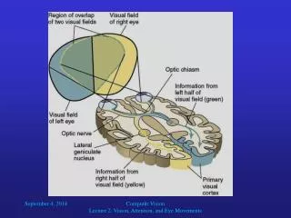
Contralateral processing • Opposite side processing is due to the optic chiasma
Nerve fibers bringing information from the right half of each visual field converge at the optic chiasma and pass to the left side of the brain • Nerve fibers bringing information from the left half of each visual field converge at the optic chiasma and pass to the right half of the brain • Information eventually ends in the visual cortex of the brain and information is shared to form a complete visual image • Image received by visual cortex is both inverted and reversed • Brain must correct in the cerebral cortex
Process of two sides of the brain working together can be illustrated by the abnormal perceptions of patients with brain lesions • Patients with right side brain lesions, when looking at an object from above, does not recognize that object and will deny what it is • Patients with left brain lesions can describe the function of the object but cannot come up with the name of the object • It takes both sides of the brain working together to have correct “vision” which is able to recognize an object and understand what it is
Vision 2020 • Joint initiative of the World Health Organization and the International Agency for the Prevention of Blindness whose goal it is to eliminate avoidable blindness worldwide by 2020 • Aim is to give every one in the world the right to sight
Videos • How the eye works • Laser surgery
Structure of the ear pinna Oval window Round window
How sound is perceived by the ear • Outer ear catches sound waves (vibrations of air molecules) • Sound waves travel down auditory/ear canal and cause the tympanic membrane (eardrum) to move back and forth slightly • Bones of the ear – malleus, incus, and stapes – receive vibrations from the tympanic membrane and multiply them approximately 20 times • Stapes strikes the oval window causing it to vibrate
Vibration is passed to the fluid of the cochlea which causes hair cells within the cochlea to vibrate • These receptors release a chemical message across a synapse to the sensory neuron of the auditory nerve and is carried to the brain • The wave in the fluid of the cochlea dissapates as it reaches the round window
Loud noises cause the fluid to vibrate to a higher degree and the hair cells bend even more • Brain interprets this as higher volume • Pitch is a function of sound wave frequency • Short, high-frequency waves produce high-pitched sound • Long, low-frequency waves produce low-pitched sound • Sound which is sensed by the brain is processes in the auditory area of the cerbral cortex
Videos • Sense of hearing • Cochlear implant