Download
1 / 28
280 likes | 302 Vues
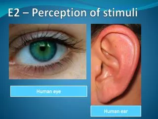
Explore the mechanisms and functions of sensory receptors such as photoreceptors, cones, rods, and bipolar cells in the human body, along with a detailed overview of the eye structure and its functions.

E N D
Sensory Receptors • Receptors in the nervous system the are sensitive to changes in the environment around the cells. • Humans have the 4 following types of specialized receptors • Mechanoreceptors • Respond to mechanical forces and movements • Chemoreceptors • Respond to chemical substances • Thermoreceptors • Respond to temperature changes • Photoreceptors • Respond to light
Smell • Detection of chemicals in the air is carried out by the Olfactory receptors. • These cells have cilia that project into the nasal cavity and their membrane contains odorant receptor molecules which assist in the sense of smell • Different organisms have differing amounts of odorant receptors • Mice have over a thousand different receptors which allows them to have a very keen sense of smell • Humans are severely lacking in odorant receptors which provides an imprecise sense of smell
Eye Functions • 1. sclera = opaque (usually white), fibrous, protective layer of the eye containing collagen and elastic fibers • 2. cornea = transparent front part of the eye that covers the iris, pupil, and anterior chamber,providingmost of an eye's optical power • 3. conjunctiva = membrane that covers the sclera (white part of the eye) and lines the inside of the eyelids • 4. eyelid - outside flesh covering of the eye. Protects from damage • 5. choroid = vascular layer of the eye lying between the retina and the sclera. The choroid provides oxygen and nourishment to the outer layers of the retina
6. aqueous humor = thick, watery substance in the eye • 7. pupil = variable-sized, black circular opening in the center of the iris that regulates the amount of light that enters the eye • 8. lens = transparent, biconvex structure in the eye that, along with the cornea, helps to refract light to focus on the retina • 9. iris = colored part of the eye • 10. vitreous humour = the clear aqueous solution that fills the space between the lens and the retina • 11. fovea = responsible for sharp central vision • 12. optic nerve = transmits visual information from the retina to the brain • 13. blind spot = the specific region of the retina where the optic nerve and blood vessels pass through to connect to the back of the eye • 14. retina = thin layer of neural cells that lines the back of the eyeball
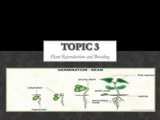
Photoreceptors • Light enters the eye through the cornea and the lens which focuses it onto the retina. • The retina is a thin layer of light-sensitive tissue at the back of the eye. • The retina involves two types of photoreceptors • Rods and Cones • Many nocturnal animals only have rods and cannot distinguish color
Rods and Cones • Rods are very sensitive to light. But not color • When exposed to bright light “bleaching” of rods can occur • Cones are able to absorb different ranges of wavelengths of light
Cones Continued • There are three types of cones in the human eye. • Red, Green, Blue • The relative stimulation of each of the three cone types allows for interpretation of color • Color vision is only in bright light and tends to fade in dim light
Bipolar cells • Rods and cones synapse with neurons called bipolar cells • Bipolar cells send the electrical impulses from the rods and cones to the Ganglion cells
Neurons and action potentials • If a rod or cone cell is not stimulated by light they will depolarize and release an INHIBITORY Neurotransmitter onto a bipolar cell. • This causes that bipolar cell to become hyperpolarized and not transmit impulses to its associated retinal ganglion cell. • When light is absorbed by a rod or cone cell it becomes hyperpolarized and stops sending that inhibitory neurotransmitter to the bipolar cell • This then allows the bipolar cell to depolarize, activating the adjacent ganglion cell. • Groups of rods send signals to the brain via a single bipolar cell • Grainy Picture • Cones send signals to the brain via their own individual bipolar cell • High Definition vision!!
Ganglion Cells • Ganglion cells have cell bodies in the retina and synapse with bipolar cells • Ganglion cells pass across the front of the retina to form the “blind spot” • The “Blind spot” is actually where the ganglion cells combine to form the Optic Nerve
The Human Ear Transmit pressure waves into auditory sounds Brain interprets the information from the receptors
Outer Ear Pinna
Middle Ear Eardrum 3 smallest bones in the body (malleus, Incus and Stapes)
Inner Ear Does both auditory and balance Cochlea and Vestibula or semicircular canals
Transmission of Sound Pinna collects sound waves and passes them to the eardrum The eardrum vibrates Vibration of the eardrum puts pressure on the ear bones Ear bones absorb the vibrations and amplify them Vibrations reach the stirrup and act like a piston to convert it to mechanical energy Mechanical energy is passed on to the cochlea via the oval and round windows As it moves through the cochlea it causes the hair cells to bend
Transmission continued • Hair cells are varying heights and are in a jelly-like substance • Larger sound waves vibrate more hair cells • The more hair cells activated (bent) the louder the sound • The more frequent the sound waves the higher the pitch (frequency)
Balance and Semicircular Canals • Major contributors to vestibular system (Balance) • Semicircular canals – 3 of them • Lateral – Horizontal • Detects movement on a horizontal plane – “NO NO NO!” • Superior – Anterior • Detects movement in lateral axis – “YES YES YES!!” • Posterior – • Detects movements across the midline • Moving your head shoulder to shoulder
These canals work similar to the cochlea in the ear. • Fluid moves through the canals and disrupts the sensory hairs located in the cupula.
Ampullae and cupula • Osseous ampullae are found in each of the three semicircular canals in the inner ear. • Within this ampullae there is a smaller structure called the cupula. • This small structure contains hair bundles that connect to neurons to detect any fluid movement within that semicircular canal • The summation of these senses helps organisms determine their position in the world and maintain their balance. • https://www.youtube.com/watch?v=6LmCbRPdd0Q
Alcohol • When alcohol is consumed it diffuses throughout the body • One location the alcohol effects dramatically is the semicircular canals • The alcohol causes the fluid within these cupula to become less viscous than the fluid in the canal. • This causes any movement to be interpreted as a large and exaggerated movement. • Can cause over reaction from brain and lack of balance