Download
1 / 44
541 likes | 1.7k Vues
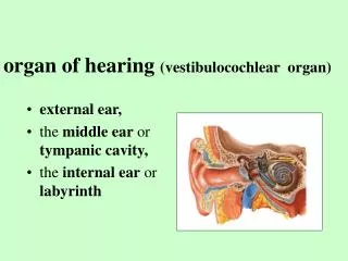
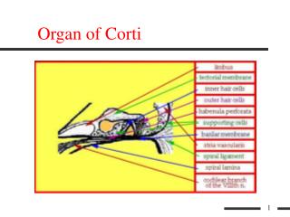
Organ of Corti. Organ of Corti (limbus). Organ of Corti (tectorial memb.). Organ of Corti (inner hair cells). Organ of Corti (outer hair cells). Organ of Corti (spriral lamina). Organ of Corti (habenula perforata). Organ of Corti (supporting cells). Organ of Corti (basilar membrane).

E N D
More on Cells • Hair Cells • Outer Hair Cells • Inner Hair Cells • Supporting Cells (modiolus to stria vascularis) • Vestibular Lip • Border Cells of Held • Inner Phalangeal Cells • Rods of Corti (Pillar Cells) • Dieters Cells • Hensons Cells • Claudius and Boettcher Cells
Cross section showing hair cells and supporting cells • Note: modiolus is to the right of slide
Inner Hair Cells • Inner Hair Cells (see previous slide) • Jug shaped • Larger than OHCs • Single Row • 3000 to 4000 in number • Innervated by both afferent and efferent fibers • IHC cilia • 50 to 70 cilia per cell in U shaped configuration. • Cilia not embedded in tectorial membrane
Outer Hair Cells • Outer Hair Cells (see previous slide) • Tube shaped • Smaller than IHCs • 3 rows except at apical end where it increases to 4 and then 5 rows • 12,000 to 13,000 in number • Innervated by both afferent and efferent fibers • OHC cilia • 58 cilia at apical end to 150 per cell at basal end in W or V shaped configuration. • Cilia embedded in tectorial membrane
Cilia • Photo showing U shaped configuration of IHC (top) and W or V shaped configuration of OHC (bottom)
Hair Cell (photo) • Outer Hair cells showing the stereocilia, reticular lamina and cuticle.
Auditory Nerve Fiber Bundle High frequency fibers: outer auditory nerve Low frequency fibers: inner auditory nerve Tonotopicity in the auditory nerve Mapping of frequency to place along the basilar membrane preserved in central auditory nervous system fiber CF 200 500 1000 Hair cells Apex: wide and loose Basilar membrane Base: narrow and tight
Central auditory system Structure Function/Description IHC to Auditory Nerve Fiber 1st synapse located in the cochlea Cochlear Nucleus 2nd synapse (1st central way station) Superior Olivary Complex 3rd synapse, Convergence of inputs from two ears, Delay line, localization Inferior Colliculi 4th synapse, multi-sensory integration Medial Geniculate Body 5th synapse, located in the thalamus Auditory Cortex 6th synapse
fiber CF 8000 Hz 4000 2000 fiber CF 200 500 1000 fiber CF 8000 Hz 4000 3000 fiber CF 200 500 700 Plasticity: flexibility, the ability to adapt to new situations. Tonotopically organized Auditory Cortex Normal Hair cell death, 1-2 kHz
Acoustic Reflex Arc • Acoustic reflex: • Contraction of the stapedius and tensor tympani muscles occurs at high intensity levels • The response is bilateral • Some functions: • Protection from intense sounds • Smoothing of the frequency response of the outer and middle ears
Acoustic Reflex Arc Midline of the brain Efferent (ipsilateral pathway) Efferent (contralateral pathway) STIMULUS Afferent