Download
1 / 1
10 likes | 190 Vues
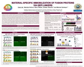
MATERIAL-SPECIFIC IMMOBILIZATION OF FUSION PROTEINS VIA GEPI LINKERS Carol Jia 1 , Marketa Hnilova 1, Banu Taktak 2 , Candan Tamerler 1,2 and Mehmet Sarikaya 1,2 1 GEMSEC, Materials Science and Engineering, University of Washington, Seattle, WA 98195, USA,

E N D
MATERIAL-SPECIFIC IMMOBILIZATION OF FUSION PROTEINS VIA GEPI LINKERS Carol Jia1, Marketa Hnilova1, Banu Taktak2, Candan Tamerler1,2 and Mehmet Sarikaya1,2 1 GEMSEC,Materials Science and Engineering, University of Washington, Seattle, WA 98195, USA, 2 MOBGAM, Molecular Biology and Genetics, Istanbul Technical University, Istanbul, 34469 Turkey, Email: markeh@u.washington.edu ABSTRACT APPROACH Peptide selection We previously selected the novel gold-binding peptides (AuBPs) from FliTrx bacterial surface library1 After five rounds of biopanning selection to enrich the population for affinitive inorganic binders, we selected and sequenced 50 clones to determine amino acid composition of randomized insertions. The binding affinities of these 50 clones were analyzed using Fluorescence Microscopy technique.2 QBPs were in silico designed based on earlier phage display selection3. The goal of this project is to immobilize enzymes and other functional biomolecules onto patterned inorganic solid surfaces. For this we apply self-assembly processes manifested by solid-binding peptides with high affinity and specificity. Current methods of binding and assembling biomolecules onto solid surfaces involve utilizing covalently bound chemical linkers. These approaches have disadvantages of limited control over molecular orientation on the substrates and limited material specificity. Also, these chemical approaches are not biocompatible. We show here that the use of combinatorially selected GEPIs, Genetically Engineered Proteins for Inorganics, could provide a more versatile alternative. Taking advantage of their high specificity and affinity to different inorganic substrates, GEPIs can be patterned onto substrates for binding with a specific biomolecular orientation. GEPIs can also be tailored for a variety of functions, including binding, linking, and producing bi-functional units.1 We previously demonstrated the utility of quartz-binding and gold-binding GEPI’s (QBP’s and AuBP’s) as highly specific biomolecular linkers.2,3 Here we first use bi-functional peptides containing both gold- and quartz-binding sequences (AuBP-QBP) and create gold patterns using soft lithography and self assembly techniques. Maltose-binding protein genetically fused with either AuBP’s or QBP’s are then introduced onto the patterned substrate to demonstrate the specificity of their binding via GEPI linker. As evidenced by a variety of experimental techniques, e.g. fluorescent microscopy (FM) and dark field microscopy (DF), we successfully immobilized various nanostructures and recombinant MBP onto solid surfaces via peptide linkers. Design of Multifunctional GEPI Construct QBP-AuBP: PPPWLPYMPPWSGGGWAGAKRLVLRREE Quartz Binding Motif Linker Gold Binding Motif Genetically Engineered MBP-GEPI Fusion Protein QBP2 Linker Peptide Synthesis: We designed novel bi-functional peptide containing both gold- and quartz- binding motifs. QBP-AuBP bi-functional peptide was synthesized using Solid Phase Peptide Synthesis (SPPS) technique. Linker AuBP1 -PGPGPG- -LPDWWPPPQLYH -SGGG- -WAGKRLVLREE mal E lacZ MCS MOTIVATION Clone Genetically Engineered MBP-GEPI Fusion constructs: Maltose-binding protein (MBP) was genetically fused to inorganic-binding peptides (e.g., AuBPs and QBPs) as follows: Specific nanobiotechnology applications (e.g. hybrid bottom-up-assembly, biofabrication, cell immobilization and biosensing applications) require selective adsorption of receptor molecules to various inorganic systems. Ideally the coupling onto an inorganic surface should be highly specific, highly ordered, reversible and biology-friendly. A novel alternative to current chemical coupling may be the utility of combinatorially-selected inorganic-binding peptides as molecular linkers.2 MBP-WT QBP-MBP AuBP-MBP Expression pMAL Marker Recombinant GEPI-MBP S(G)3 (PG)3 S(G)3 (PG)3 RESULTS FUFURE DIRECTIONS RESULTS Simultaneous Immobilization of Various Enzymes MBP-GEPI Ink in Micro-contact Printing MBP-GEPI Self Assembly on Patterned Surfaces Colorimetric Detection of Enzymatic Cascade Reactions: 1) Creation of Patterned Surfaces via Bifunctional Peptides 1) MBP-QBP Fusion Protein on Silica Glass : GEPI1- Enzyme1 (e.g. glucose oxidase) Color change + substrate e.g. ABTS ABTS+ (Absorbance) AuBP NC : Enzyme2 (e.g. peroxidase) MBP NC : Substrate1 (glucose) PDMS PDMS : Substrate2 / Product 1 (H2O2) : QBP-MBP (Printing) : AuBP-QBP (Printing) GLASS 10 10 m : BSA (Blocking) GLASS : Product2 (H2O) 10 m QBP-PPP-AuBP QBP GLASS : AuNP (Self Assembly) MBP-SGGG-QBP MBP-PGPGPG-QBP : anti-MBP-Alexa GLASS Y Y Y Y Y Y Y Y GLASS Fig.: Bifunctional QBP-AuBP and QBP and AuBP proteins (200µg/ml) were incubated on PDMS stamps for 15 min, excess proteins were removed and the PDMS was dried with nitrogen, then the stamps were incubated with silica glass surface for 15 min. After stamping, the surfaces were washed and dried with nitrogen. The immobilized proteins were detected by gold nanoparticles (4.5x10¹⁰ particles/ml) using Dark Field Microscopy technique. Fig.: Recombinant fusion QBP-MBP and MBP-WT proteins (1mg/ml) were incubated on PDMS stamps for 15 min, the excess proteins were removed and the PDMS dried with nitrogen. Then stamps were incubated with silica glass surface for 15 min. After stamping, the surfaces were washed and dried with nitrogen. After blocking the surface with BSA (1 mg/ml) for 1 hr and washing the surfaces, the immobilized proteins were detected by anti-MBP monoclonal antibody labeled with Alexa dye (1ug/ml) using Fluorescent Microscopy technique. GLASS CONCLUSIONS 2) Self Assembly of MBP-GEPI on Patterned Surface • Previously we selected gold- and quartz-binding peptide (AuBP and QBP) sequences from combinatorial peptide libraries. We demonstrated the utility of combinatorial selected inorganic-binding peptides, such as gold- and quartz-binding peptides (AuBPs and QBPs), as highly specific biomolecular linkers to direct immobilization of variety of inorganic nanostructures • We synthesized bifunctional QBP-AuBP, and demonstrated its efficacy in the assembly of gold nanoparticles, resulting gold patterned surfaces were characterized using dark field optical and atomic force microscopies. • We genetically fused AuBP and QBP GEPIs to MBP protein and expressed the proteins in bacterial cells • As evidenced by a variety of experimental techniques, e.g. fluorescent microscopy (FM) we successfully immobilized various nanostructures and recombinant MBP-GEPI onto solid surfaces via these specific peptide linkers 2) MBP-AuBP Fusion Protein on Gold Surface AFM scan of Micropatterned AuNP MBP-WT DF : MBP-AuBP(Self Assembly) GLASS PDMS MBP NC Micro-patterned AuNP : AuBP-MBP (Printing) 10m Au : BSA (Blocking) GLASS MBP-WT FM : BSA (Blocking) 10 m MBP-SGGG-AuBP MBP-PGPGPG-AuBP Au : anti-MBP-Alexa GLASS Y Y : anti-MBP-Alexa Y Y Y Y Y Y Y Y Y Y Y Y Y Y MBP GEPI fusion protein immobilized on gold, detected by anti-MBP labeled with Alexa dye Y Fig.: Recombinant fusion AuBP-MBP and MBP-WT proteins (1mg/ml) were incubated on PDMS stamps for 15 min, the excess proteins were removed and the PDMS dried with nitrogen. Then the stamps were incubated with gold surface for 15 min. After stamping, the surfaces were washed and dried with nitrogen. After blocking the surface with BSA (1 mg/ml) for 1 hr and washing the surfaces, the immobilized proteins were detected by anti-MBP monoclonal antibody labeled with Alexa dye (1ug/ml) using Fluorescent Microscopy technique. References Au Fig.: Recombinant fusion AuBP-MBP protein (1mg/ml) was incubated on patterned substrate for 2 hr, the excess proteins were removed and the substrate washed and dried with nitrogen. After blocking the surface with BSA (1 mg/ml) for 1 hr and washing and drying the surfaces, the immobilized proteins were detected by anti-MBP monoclonal antibody labeled with Alexa dye (1ug/ml) using Fluorescent Microscopy technique. GLASS Z Lu et al., Biotechnology, 13, 366 (1995) 2. Hnilova et al., Langmuir, 24, 12440 (2008) 3. Oren et al., Bioinformatics, 23, 2816, (2007) The research is supported by NSF-MRSEC Program through the University of Washington GEMSEC (DMR 0520567), NSF-BioMat, and IRES Programs, and TUBITAK (TR). MBP-SG- AuBP DF MBP-SG- AuBP FM