
Circulatory system
Discover the structure and functions of the circulatory system, including blood vessels, the heart, and lymphatics. Learn about the different types of arteries and veins, the microcirculatory bed, and the role of the lymphatic system. Explore the anatomy of the heart and its various chambers. Gain a deeper understanding of the immune and hemopoietic organs, including the red and yellow bone marrow, thymus, spleen, and lymph nodes. Dive into the world of endocrine regulation and hormone activity.

Circulatory system
E N D
Presentation Transcript
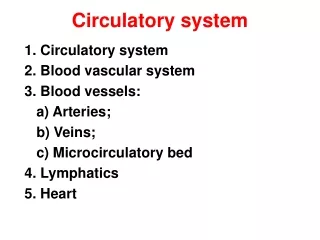
Circulatory system 1. Circulatory system 2. Blood vascular system 3. Blood vessels: a) Arteries; b) Veins; c) Microcirculatory bed 4. Lymphatics 5. Heart
Scheme of vessel’s wall1. Intima:a) endothelium; b) subendothelial layer2. Media (sm. myocytes+ fibers) 3. Adventitia (loose con. tissue)
1. Intima:a) Endothelium; b) Subendothelial layer; c) Internal elastic lamina2. Media(sm. myocytes+ fibers) 3. Adventitia(loose con. tissue+ vasavasorum)
Elastic artery1. Large endothelial cells2. Subendothelial layer (myointimal cells)3. Elastic fibers predominate
Muscular artery1. Thin intima 2. Myocytes predominate 3. Internal elastic lamina well defined
Artery-vein1. Thin wall2. Bigger diameter (irregular shape)3. Collagen fibers (laminas) 4. Adventitia thick5. Walves
Anastomoses • AVA functions are the next: 1.Regulation (correction) of the blood pressure. 2.Blood supplying of organs. 3.Venous blood saturation with oxygen. 4.Blood withdrawing from the depot. 5.Regulation of the tissues fluid passage to the venous bed. Types: typical+ nontypical
HEART1. Endocardium (endothelium, subendothelial layer, fibro-muscularand external connective tissue layer)2. Myocardium(contractile, conductive,secretory myocytes)3. Epicardium4. Pericardium
Immune and hemopoietic organs Hemopoietic organs classification. Сommon featuresof hemopoietic organs Red bone marrow Yellow and mucous bone marrow Involution of thymus Spleen Lymph nodes
Hemopoietic organs Central -antigenindependent • Red bone marrow • Thymus Peripheral -antigendependent • Spleen • Lymph nodes • MALT • Appendices • Peyer’s patches
General features 3. Functions • Hematopoiesis • Deposition • Protective • Elimination 1. Origin - mesenchyme 2. Structure a. Stroma Reticular tissue Sinusoids Macrophages b. Parenchyma Myeloid tissue Lymphoid tissue
Stromal cellsParenchyma • Reticular cells hemopoietic islets: • Osteogenic cells erythropoietic • Adipocytes trombopoietic • Adventitial cells granulocytopoietic • Endothelial cells agranulocytopoieti • Macrophages
Thymus Functions 1.Antigen-independent proliferation of T-lymphocytes2. Endocrine Stroma -- epithelio-reticular tissueParenchyma-lymphoid tissue
MFU – lobule (cortex, medulla)Hematothymic barrier1. Endothelium2. Basement membrane3. Epithelio-reticulocyte4 Perivascular space with macrophages
Endocrine system 1. Neuro-endocrine-immune regulation. 2. Hormones and mechanisms of their activity. 3. Endocrine organs classification. 4. Central endocrine organs: а) hypothalamus; б) pituitary; в) epiphysis. 5.Peripheral endocrine glands: thyroid, parathyroid
Neuro-endocrine-immune regulation Nerve system Liberins and statins cytokins neurotransmitters hormones Immune system Endocrine system corticoateroids
Endocrine glands peculiarities1. Stroma + parenchyma.2. Endocrine cells.3. Ductless glands.4. Well developed microcirculatory bed with fenestrated capillaries • Hormones • 1.Proteins derivatives • 2.Especially active • 3.Selective influence on “target-cells” • Mechanisms of hormones activity1. Cell membrane permeability changes • 2. Activation of intracellular processes3. Influence on the chromosomes
Parenchymal unites of endocrine glands1.Lobule2.Trabecula3.Follicle Central organs:hypothalamus, hypophysis, epiphysis Peripheral glands:1. Endocrine : а) pituitarydependent (thyroid; adrenal cortex); b) pituitaryindependent (parathyroid). 2. Mixed (mammary, testes, ovaries, Langerhans islets) 3. Diffuse endocrine system
Anterior hypothalamus(large cholinergic cells): а) supraoptical nuclei – antidiuretic hormone;b) paraventricular nuclei - oxytocin. • Middle hypothalamus -5 pares of nuclei • (small adrenergic cells) • (ventromedial, dorsomedial, arcuate, suprahiasmatic та preoptic zone)- relising factors (liberins and statins)
Hypothalamo-adenohypophysial systemHypothalamo-nerohypophysial system
Adenopituitary:trabecules1. Acidophils– somatotrophs- andmammotrophs.2.Basophils – thyrotrophs and gonadotrophs.3. Intermediate cells – adrenocorticotropocytes.1. Melanotrophs 2. LipotrophsNeuropituitary:pituicytes + axovasal synapses (Herrings bodies)
Epiphysis(pineal gland)lobulespinealocytes: light and dark + astrocytes Corpora arenacea Parathyrocytes cords Trabecules
Parathyroid glandsstructural unit – cord(chief cells and oxyphilic cells)
Cortex(adrenocorticocytes)Zona glomerulosaZona fasciculatazona reticulosa Medulla Epinephrocytes and norepinephrocytes


