Download
1 / 55
580 likes | 1.09k Vues
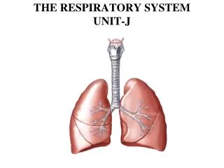
THE RESPIRATORY SYSTEM UNIT-J. Objectives: 1. Describe the structure of the Respiratory System 2. Analyze the function of the Respiratory System 3. Identify characteristics and treatment of common Respiratory disorders. NOSE or MOUTH

E N D
Objectives: 1. Describe the structure of the Respiratory System 2. Analyze the function of the Respiratory System 3. Identify characteristics and treatment of common Respiratory disorders
NOSE or MOUTH PHARYNX LARYNX TRACHEA RT. or LT. BRONCHUS ALVEOLI BLOOD Pathway of Air into Lungs
Alveoli Apex Bronchi Bronchioles Cilia Coughing Epiglottis Expiration/exhalation Hiccups Inhalation/inspiration Larynx Lobes Lungs Medulla oblongata Nose (nasal cavity) Phrenic nerve Pleura Pleural cavity Respiration Sinuses Sneezing Surfactant Trachea Ventilation Yawning TERMINOLOGY
EPISTAXIS HYPOXIA INFLUENZA INTERCOSTAL LARYNGITIS NARE PHARYNGITIS PHARYNGOSPASM APNEA ASTHMA BRONCHITIS BRONCHIECTASIS COMMON COLD DYSPNEA E.E.N.T EMPHYSEMA DISORDERS AND RELATED TERMINOLOGY
PLEUROCENTESIS RHINIRRHEA SUBLINGUAL TACHYPNEA THORACOTOMY THRACHEOSTOMY TUBERCULOSIS URI PNEUMONIA PNEUMONOLYSIS PNEUMOTHORACIS PULMANARY EDEMA RALES RHINOPLASTY DISORDERS AND RELATED TERMINOLOGY
Nasal Septum Divides nasal cavities into R and L cavities. Turbinate are three scroll- shaped bones that protrude into the nasal cavity-they increase surface area for filtering dust and dirt particles by the mucous membrane. NASAL CAVITY
They are hairs located in the nose (nasal epithelium), they filters out air and trap larger dirt particles. Cilia
The Throat Common passageway for air and food 5” long When food is swallows, the EPIGLOTTIS closes over the opening to the larynx, preventing food from entering the lungs. Pharynx
Voice production Triangular chamber below pharynx (inside the neck) Within the larynx are vocal cords (GLOTTIS) Adam’s Apple Speech is made possible by the fibrous plates contained within the cartilage of the larynx. LarynxVoice Box
Sinuses Cavities in the skull, that produce mucous for the respiratory tract, lined with mucous membrane to warm and moisten the air. • Frontal • Maxillary • Ethmoid • Sphenoid Sinuses give resonance to the voice
Is composed of a single layer of epithelial tissue with millions of tiny, thin walled sacs. SURFACTANT is a fatty substance in the lungs that prevents the alveoli from collapsing. Each alveolus surrounded by capillaries. O2 and CO2 exchange takes place between the alveoli and capillaries Alveoli
Windpipe 4 ½ inches long The walls of trachea are made more rigid by the C-shaped rings of hyaline cartilage-to keep trachea open. Lined with ciliated mucous membrane. Coughing and expectoration gets rid of dust-laden mucous. Trachea
Lower end of trachea divides into Rt. and Lt. bronchi As they enter the lungs, subdivide into bronchial tubes and bronchioles Bronchi-similar to trachea with ciliated mucous membrane and hyaline cartilage Bronchial tubes-thinner walls of smooth muscle, lined with ciliated epithelium At the end, alveolar duct and cluster of alveoli Bronchi and Bronchioles
A thin, moist slippery membrane that lines the outer surface of the lungs and the inner surface of the rib cage. Double-walled sac Space is pleural cavity-filled with pleural fluid to prevent friction. Pleura
Each lung is divided into two or three lobes. Fill thoracic cavity Upper part=apex Lower part=base Base fits snugly over diaphragm Lung tissue porous and spongy- it floats Rt. lung= larger and shorter (displaced by the liver) and has 3 lobes Lt. lung smaller (displaced by the heart)and has 2 lobes Lungs
PULMONARY VENTILATION Breathing
The part of respiration that involves air being taking into the lungs. The intercostal muscle lifts ribs outward, sternum rises and the diaphragm contracts and moves downward-this increases the volume of the lungs and air rushes in. INSPIRATION
Opposite action takes Place. Exhalation is a passive Process. EXPIRATION
A deep prolonged Breath that fills the lungs, increases oxygen within the blood. YAWNING
A deep breath followedby forceful expulsion of air –to clear lower respiratory tract. COUGHING
1 inspiration + 1 expiration = 1 respiration. Normal # of breaths an adult takes each minute-14-20. Increases with exercise, body temperature, certain diseases. Age – newborn = 40-60/min Sleep = respirations ↓ Emotion can ↑ or ↓ RESPIRATORYMOVEMENTS
HICCUPS They are a spasm of the diaphragm and spasmodic closure of the glottis- irritation to diaphragm or phrenic nerve
Air is forced through the nose to clear respiratory tract. SNEEZING
NEURAL FACTORS Respiratory center located in MEDULLA OBLONGATA. ↑ on CO2 or ↓ O2 in the blood will trigger respiratory center. PHRENIC NERVE – stimulates the diaphragm. CONTROL OF BREATHING: breathing controlled by neural and chemical factors.
Spirometer – device that measures lung capacity Tidal Volume – amount of air that moves in and out of lungs with each breath. Normal = 500 ml LUNG CAPACITY AND VOLUME
CO2 and O2 levels in the blood is sensed by the brain (respiratory center in brain). Chemoreceptor in aorta and carotid arteries sensitive to the amount of blood O2. CHEMICAL FACTORS
SINUSITIS Infection of mucous membrane that lines sinus cavities Caused by bacteria or virus Symptoms – headache or pressure, thick nasal discharge, loss of voice resonance Rx – symptomatic, surgery for chronic sinusitis RESPIRATORY DISORDERS
Contagious viral respiratory infection Indirect causes – chilling, fatigue, lack of proper food, and not enough sleep Rx – Rest, drink warm liquids and fruit juice, good nutrition Also called an Upper Respiratory Infection (URI) Hand washing – best preventative measure COMMON COLD
TUBERCULOSIS • Illegal immigration, homelessness and AIDS has caused an in US. • Tubercles (lesions) form in the lungs • Symptoms: cough, low grade fever in the afternoon, weight loss, night sweats • Diagnosis – TB skin test • If skin test positive – follow up with chest x-ray and sputum sample • Rx – antibiotic
Inflammation of larynx or voice box. Often secondary to other respiratory infections. Symptoms – sore throat, hoarseness or loss of voice, dysphasia (difficulty swallowing). LARYNGITIS
PHARYNGITIS – Red inflamed throat. REPIRATORY DISORDERS CONTINUED…
Inflammation of the lining of the lungs. Usually occurs in conjunction with pneumonia and other lung infections. Symptoms – sharp, stabbing pain when breathing, dyspnea and fever PLEURISY
Viral infection (VIRUS) causing inflammation of the mucous membrane. Fever, mucopurulent discharge, muscular pain, extreme exhaustion. Complications – pneumonia, neuritis, otitis media & pleurisy. Rx – treat the symptoms INFLUENZA (Flu)
Infection of the lung Caused by bacteria or virus. Alveoli fill with exudates (thick fluid) Symptoms – chest pain, fever, chills dyspnea. Rx – O2 and antibiotics PNEUMONIA
Inflammation of the mucous membrane of the trachea and bronchial tubes, producing excessive mucous May be acute or chronic Acute bronchitis characterized by cough, fever, substernal pain and RALES (raspy sound). Chronic bronchitis – middle or old age, cigarette smoking most common cause BRONCHITIS
Inflammatory airway obstruction Caused by allergen or psychological stress 5% of Americans have asthma Symptoms: difficulty exhaling, dyspnea, wheezing, tightness in chest Rx: anti-inflammatory drugs, inhaled bronchodilator ASTHMA
NASAL POLYPS Growths in sinus cavity, cause obstruction in air pathway Rx: surgical removal
Cause: breathing dust containing silicon dioxide over long period of time. Lungs become fibroses, reduced ability to expand. SILICOSIS
Collapsed lung due to air in the pleural cavity. PNEMOTHORAX
Insertion of a needle through the thoracic cavity and into the pleural cavity to drain fluid. THORACENTESIS
(COPD) – Describes chronic lung conditions, especially emphysema and chronic bronchitis Rx – alleviate the symptoms, decrease exposure to respiratory irritants, prevent infections, restructure activities to prevent need for O2 CHRONIC OBSTRUCTIVE PULMONARY DISEASE
Lungs fail to expand normally due to bronchial occlusion, (something that obstructs). ATELECTASIS
Alveoli becomes over dilated, lose their elasticity. May eventually rupture Air becomes trapped, can’t exhale – forced exhalation required. Reduced exchange of O2 and CO2. Dyspnea increases as disease progresses EMPHYSEMA
PERTUSSIS-Called the (Whooping cough) Symptoms: severe coughing attacks that end in a “whooping” sound, dyspnea. Prevented by a childhood Vaccine. REPIRATORY DISORDERS
An upper respiratory tract illness characterized by sore throat, low fever, & a pseudomembrane on the tonsils, pharynx,&/or nasal cavity.Rare infectious disease. Caused by Corynebacterium diphtheria. Prevented by childhood vaccine DIPTHERIA
Dilation of a bronchus caused by inflammation Heavy pus secretion BRONCHIECTASIS
Inflammation of nasal mucosa with increased secretion Caused by virus allergens RHINITIS