Laser Applications
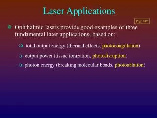
Laser Applications. Page 149. Ophthalmic lasers provide good examples of three fundamental laser applications, based on: total output energy (thermal effects, photocoagulation ) output power (tissue ionization, photodisruption ) photon energy (breaking molecular bonds, photoablation ).

Laser Applications
E N D
Presentation Transcript
Laser Applications Page 149 • Ophthalmic lasers provide good examples of three fundamental laser applications, based on: • total output energy (thermal effects, photocoagulation) • output power (tissue ionization, photodisruption) • photon energy (breaking molecular bonds, photoablation)
Ophthalmic Lasers Page 149 • Ophthalmic lasers make use of the spectral transmission properties of the eye • The cornea transmits all visible wavelengths plus infrared • The highly collimated laser beam allows precise focusing on target tissue
Photocoagulation • Most common (and first used) ophthalmic laser procedure • Laser “seals” hemorrhaging vessels (usually retinal vessels) • Also used to destroy areas of neovascularization, and to obliterate retinal or subretinal tumors
Photocoagulation - Rationale • Photocoagulation works byheating “target” tissueenough e.g. to seal the hemorrhaging vessels, or destroy a tumor, whilesparing adjacent tissue • The heating process relies ontotal energy deliveredto the target site. Continuous wave lasers are adequate for this purpose
Lasers used for Photocoagulation Page 152 • Continuous waveArgon or Kryptonlaser; exposure controlled by an external shutter • Nd-YAG pulsed laserset in “quasi-continuous” mode (long duration, e.g. 1 sec) pulses.
Thermal Effects & Ocular Chromophores Page 150 • To heat target tissue, photons must be absorbed by the tissue and initiate a chemical change • Chromophore: target molecule (or molecular region) that will absorb a photon of a particular energy level • After absorption by chromophores, photons maybreak molecular bonds, ionize the molecule, or causemolecular excitation.
Ar 515 Kr 647 Ar 488 Kr 568 Nd-YAG 1,064 nm melanin HbO2 Deoxy-Hb xanthophyll Absorption Spectra of Important Ocular Chromophores 104 103 Extinction Coefficient (cm-1) 102 10 1 400 500 600 700 800 900 1000 1100 Fig 92, p 150 Wavelength (nm)
Retinal Photocoagulation and Chromophores A good example to demonstrate the use of chromophores photocoagulation of macular microaneurysms (diabetes)
Macula lutea The Macula lutea (yellow spot) Color greatly exaggerated
Photocoagulation of Macular Microaneurysms • Laser energy must be absorbed by the blood in the microaneurysms • Must target chromophores in blood look at absorption spectrum of hemoglobin
Ar 515 Kr 647 Ar 488 Kr 568 Nd-YAG 1,064 nm melanin 560 – 580nm range optimum HbO2 Deoxy-Hb xanthophyll Absorption Spectra of Important Ocular Chromophores 104 103 Extinction Coefficient (cm-1) 102 10 1 400 500 600 700 800 900 1000 1100 Wavelength (nm)
Photocoagulation of Macular Microaneurysms Avoiding surrounding, non-target chromophores • Xanthophyll (macular pigment) is also a chromophore • Excessive absorption of energy by xanthophyll will cause macular/photoreceptor damage • Xanthophyll absorbs shorter wavelengths (blue)
Ar 515 Kr 647 Ar 488 Kr 568 Nd-YAG 1,064 nm melanin Avoid ’s below 525 nm HbO2 Deoxy-Hb xanthophyll Absorption Spectra of Important Ocular Chromophores 104 103 Extinction Coefficient (cm-1) 102 10 1 400 500 600 700 800 900 1000 1100 Wavelength (nm)
Retinal Photocoagulation and Chromophores Other Aspects • Shorter wavelengths: • have higher photon energy and are more efficient at heating target chromophores • do not penetrate as far through tissue • elicit greater Rayleigh scatter • Longer wavelengths: • are less efficient at heating target chromophores • penetrate tissue to greater depths and may damage underlying tissue • elicit less Rayleigh scatter
Photocoagulation: pre-surgical appearance Severe proliferative vitreoretinopathy - new vessel and fibrous tissue growth prior to laser surgery
Following photocoagulation After panretinal laser photocoagulation, abnormal vessels gone; fibrous tissue remains
Page 156 Photodisruption • Optical breakdown of target tissue by ionization • Results in “plasma” formation as electrons dissociate from their atoms • Plasma formation both audible and visible as spark • Local temperature elevations to 10,000O C • Requires high power laser (PULSED)
Photodisruption • A Q-switched Nd-YAG laser often used: • causes photodisruption mainly by focal heating thermionic emission (from linearly absorbing chromophores) • Higher power mode-locked Nd-YAG laser (20-30 psec pulses): • ionizes by multiphoton absorption • each 1,064 nm photon carries 1.17 eV of energy • because atoms typically must absorb > 10 eV to ionize need “multi”-photon absorption for ionization
Ophthalmic Photodisruption Page 158 • Ophthalmic surgical procedures that utilize photodisruption include: • Posterior capsulotomy to open up an opacified lens capsule resulting from prior extracapsular cataract surgery (small incision made in lens capsule and lens contents extracted by vacuum)
(Months) Post-op. Appearance with Implants PMMA Implant(a) regenerated posterior capsular cells impair vision(b) scatter light Silicone Implant(a) clear posterior capsule(b) no axial scatter Soft acrylic Implant(a) some regenerated cells toward periphery (b) minimal scatter From: Hayashi: Arch Ophthalmol, Volume 116(12).December 1998.1579-1582
Implant Lens Posterior Capsulotomy with Nd-YAG Laser
Central post-capsular cells Posterior Capsulotomy with Nd-YAG Laser Implant Clip Before After
Central post-capsular cells Central post-capsular region clear Posterior Capsulotomy with Nd-YAG Laser Implant Clip Before After
Page 159 Photoablation • Use of high energy photons to directly break molecular bonds with sufficient photon energy, get very clean removal of tissue • Utilize excimer laser (excimer= excited dimer): • e.g Ar*F (argon fluoride) laser. Ar*F exists normally in a metastable state. • excimer decay produces Ar + F + high energy UV photon (193 nm 6.4 eV energy) • dissociation products rapidly reform original reactants • Kr*F not quite as effective: 248 nm 5.0 eV energy) • Excimer effect on the cornea ablative photodecomposition
Corneal Photoablation • Both PRK and LASIK utilize photoablation • Ablation precise in terms of surface coverage and ablation depth: • can therefore sculpt the cornea to a very high degree of accuracy • myopia radially symmetrical flattening of the central cornea (decrease corneal power) • hyperopia relative steepening of central cornea (increase corneal power)
Rabbit cornea showing clean region of ablation through half of corneal thickness
Typical Size of Region Ablated by PRK PRK: epithelium scraped off and photoablation applied directly to the central 5 – 7 mm corneal region
LASIK: Laser-ASsisted In-situ Keratomilieusis Excimer laser sculpting of the underlying corneal stroma Source: Horn Eye Centers, IL
PRACTICE PROBLEMS
Practice Problem 32 Photocoagulation of microaneurysms (localized capillary swelling) at the macula requires careful selection of laser wavelength. The most appropriate wavelength would: • be as short as possible, but not so short that it is absorbed by the cornea • be absorbed by the macular pigment xanthophyll, to avoid damage to the underlying choroid • be absorbed by both hemoglobin and xanthophyll (target chromophores) • be absorbed by hemoglobin (target chromophore), while not being absorbed by xanthophyll Physical Optics PS – Questions 51-54
Practice Problem K Red laser output would be the most suitable wavelength for photocoagulation when : • the lesion does not involve the macula • the lesion is vascular in origin • the target tissue is a sub-retinal lesion • it is important that there is absolutely minimal thermal transfer to adjacent tissues
Practice Problem 33 Photoablation is the laser technique used in PRK (photorefractive keratectomy) and LASIK (Laser-assisted in situ keratomilieusis). The excimer laser emits: • extremely short-duration, high power pulses that ionize corneal tissue • high-energy UV photons that break molecular bonds in corneal tissue • a specifically timed dose of laser energy that causes highly localized heating of tissue and a visible spark • a continuous wave output to cause focal coagulation of stromal tissue, while sparing the epithelium Physical Optics PS – Question 56
Practice Problem L Photodisruption occurs through a process involving : • tissue ionization • breaking of molecular bonds • thermal transfer • injecting a photoactive dye which, after reaching the target tissue, is chemically activated by a specific laser wavelength
Practice Problem M Photorefractive keratectomy is most efficient when the laser output has : • sufficiently short pulse duration to maximize pulse power and achieve corneal ablation • sufficiently high photon energy to achieve corneal ablation • a wavelength that is minimally absorbed by organic molecules, to prevent excessive thermal transfer during corneal ablation • sufficiently high pulse power to achieve plasma formation, which both causes corneal ablation and shields underlying structures from inadvertent damage
Practice Problem N Brewster windows operate in continuous beam lasers to : • produce a plane-polarized output beam by only allowing stimulated emission in one plane based on the principle that the exciting and (stimulated) emitted photon have an identical state of polarization • produce a plane-polarized output beam by excluding all other polarization states based on polarization by reflection • reduce reflected glare and allow a more pure axial beam to be amplified through the system • only allow "cavity modes" to oscillate through the system, based on the principle of "beat frequencies."