Download
1 / 18
180 likes | 253 Vues
This guide covers essential aspects of intubation including indications, equipment required, patient preparation, communication strategies, and post-intubation procedures to ensure successful respiratory services.

E N D
Intubation Assist Respiratory Services Oct. 2009
Intubation Assist • Indications for intubation • Endotracheal tube sizes • Equipment required for ETT intubation • Equipment preparation • Patient Preparation • Communication • Post Intubation • Rapid Sequence Intubation
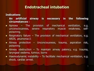
Indications for Intubation • To prevent or relieve upper airway obstruction. • To protect the airway from gross aspiration. • To facilitate tracheal suctioning. • To provide a closed system to deliver positive pressure ventilation.
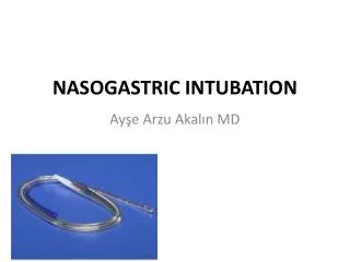
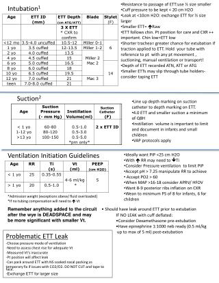
Equipment Required for ETT Intubation • Typical Oral ETT Sizing: Adult Female 7.0-8.0 O.D with approximate insertion depth of 20-22 cm. Adult Male 7.5-8.5 O.D with approximate insertion depth of 22-24 cm. ** EVAC ETT’s outer diameter is, on average, 0.8 mm larger than a standard ETT, so one should consider using an EVAC ETT one half size smaller than usual.** Evac Tubes are available in sizes 6-9 O.D.
ETT Sizing - Pediatrics • Standard ETT for pediatric patients is 4.0-6.5 O.D • Predicted size for Pediatric ETT= age/4 + 4 Age Tube Size Tube length(incisors to tip) 18 mo 3.5-4.5 11-13 3 yrs 4.5-5.0 12-14 5 yrs 4.5-5.0 13-15 6 yrs 5.5-6.0 14-16 8 yrs 6.0-6.5 15-17 12 yrs 6.0-7.0 17-19 16 yrs 6.5-7.0 18-20
ETT Sizing - Neonates Birth weight (g) Gestational Age (Wks) ETT Size (mm) Tube Length(cm) < 1000 Below 28 2.5 9-11 1000- 2000 28-34 3.0 9-11 2000 - 3000 34-38 3.5 10-12 > 3000 > 38 3.5 - 4.0 11-12
Equipment Required for Intubation • Laryngoscope handle • Laryngoscope blade – Mac 3, Mac 4 (curved blades) Miller 4 (straight) • Magill forceps • Stylet • ETT securing device • ETT tubes in various sizes • Water soluble lubricant • Xylocaine spray • 10 cc syringe • Assorted size of oral or nasopharyngeal airways • Suction equipment including yaunker and suction catheters • Bag-valve-mask • ETCo2 monitoring device or esophageal intubation detecting device (EDD)
Equipment Cont’d • Only required equipment should be taken into the patient’s room. This will prevent contamination and need for disinfection or disposal of unused supplies. • It is helpful to have another staff member remain outside the patient room to obtain extra equipment without contaminating a whole intubation kit.
Bag-Valve-Mask (BVM) • The patient will require pre-oxygenation with 100% O2 and hyperventilation (except with Rapid Sequence Intubation) via a BVM. • It is vital to ensure that a filter is placed between the mask and bagging unit to minimize risk of exposure to infection.
Equipment Preparation • Prior to intubation all required equipment should be prepared for use: • Check cuff on ETT • Insert stylet • Lubricate ETT • Check light on laryngoscope blade • Set up End tidal CO2 or open EDD • Check suction set up • Prepare ETT securing device
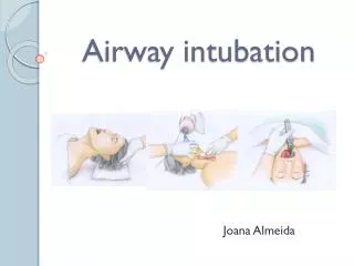
Patient Preparation Patient position is vital in preparing for intubation • Ensure bed is at appropriate height for person intubating. • Patient should be positioned as close to the head of the bed as possible. • Patient’s head should be positioned such that the mouth, pharynx and larynx are aligned. This is achieved by combining moderate cervical flexion and extension of the atlanto-occipital joint. • Placement of a small pillow or towel under the head can help achieve this position.
Team Communication Communication amongst team members is very important during intubation • The person assisting intubation should ask the person performing it what their back up plan is if the first attempt is unsuccessful. • The person assisting can then have alternative airway adjuncts ready to be used or have extra personnel on stand by to assist.
Communication • During the intubation, the person assisting should ask the intubator if he/she can visualize the vocal cords and if they require laryngeal pressure. • Laryngeal pressure is often referred to as “BURP” which stands for: Backward, Upward, Rightward, Pressure. • This pressure is applied to the thyroid cartilage which in turn presses against the larynx and helps bring it into full view.
After Placement of ETT Once the ETT is in place assessment of correct position must begin: • Attach EDD or ETCO2 monitor. • Auscultate over the stomach first, then the lungs, checking for bilateral breath sounds. • Watch for misting of the ETT. • Once placement has been achieved, check the tube position and secure tube into place. • ETT placement should then be confirmed with chest X-ray.
Rapid Sequence Intubation (RSI) • In a RSI, the patient is pre-oxygenated and rapidly rendered unconscious and paralyzed within 45-60 seconds to facilitate emergent ETT intubation without the use of bag valve mask ventilation to minimize the risk of aspiration.
RSI Procedure • Pre-oxygenate the patient with 100%O2 for 3-5 min. by placing the BVM over the patient’s face, creating a tight seal and allowing the patient to breathe on their own. • This will wash out nitrogen and establish an oxygen reservoir. This reservoir will allow for several minutes of apnea without arterial desaturation. * A healthy adult can maintain an SpO2 >90% for 8 min. of apnea with pre-oxygenation. * Young children, adults with cardiorespiratory disease, obese patients and pregnant women may desaturate to <90% in less than 3 min. • BVM ventilation should only be provided if the patient’s SpO2 falls below 90%.
RSI Procedure • The patient’s airway can be protected from aspiration by avoiding BVM ventilation and applying cricoid pressure. • Cricoid pressure (Sellick’s maneuver) is achieved by using the thumb and index or middle finger to apply firm downward pressure on the cricoid cartilage. This will cause compression of the esophagus against the vertebrate and prevent regurgitation of gastric contents into the larynx and pharynx. • Cricoid pressure should only be applied after the patient is rendered unconscious and should not be released until ETT placement has been confirmed.
Airway Adjuncts A variety of airway adjuncts are available if conventional intubation cannot be achieved • Staff must make themselves aware of the various devices available in their hospital, how to use them and where these pieces of equipment are located. • Examples of the adjuncts are: 1. Bougie 5. LMA 2. Glidescope 6.. Fastrach LMA 3. Lighted Stylet 7. Bronchoscope 4. McCoy Blade 8. Surgical airway equipment