Download
1 / 61
610 likes | 635 Vues
This chapter discusses the primary functions of the blood, its physical characteristics, major components, and the two major fluid systems - the cardiovascular system and the lymphatic system.

E N D
Chapter 19: The Blood BIO 211 Lecture Instructor: Dr. Gollwitzer
Today in class we will discuss: • The primary functions of the blood • List the physical characteristics and major components of the blood • Plasma • Describe the composition and functions of plasma • Describe the roles of various plasma proteins • Red Blood Cells (RBCs) • List the characteristics and functions of RBCs • Describe the structure and function of hemoglobin • Explain the basis for ABO and Rh blood types and the cause of incompatibilities
2 Major Fluid (Circulatory) Systems • Cardiovascular system (CVS) • Circulating fluid = blood (Chapter 19) • Pump = heart (Chapter 20) • Conducting “pipes” = blood vessels (Chapter 21) • Lymphatic system (Chapter 22) • Interconnected and interdependent with CVS • CVS (bloodstream) fluid tissues fluid lymphatic vessels CVS • CVS assists lymphatic system (defense system) – blood carries lymphatic cells, antibodies, cytokines, etc.
Blood • Specialized fluid connective tissue • Contains • Cells in fluid matrix • Proteins • Functions • Transport of dissolved substances • Regulation of pH and ion composition • Restriction of fluid loss at injury sites • Defense against toxins and pathogens • Stabilization of body temperature
Functions: Transportation • Dissolved gases • Oxygen (O2) from lungs to tissues • Carbon dioxide (CO2) from tissues to lungs • Nutrients • Absorbed from digestive tract, adipose tissue, liver • Hormones • From endocrine glands to target cells • Metabolic wastes • Absorbs and carries from tissue cells to liver • Immune system cells • Defend tissues from infection and disease
Functions: Regulation of pH and Ion Composition of Interstitial Fluids • Occurs via diffusion between blood and interstitial fluids • Eliminates local deficiencies or excesses of ions • e.g., increases/decreases calcium or potassium • Absorbs and neutralizes acids • e.g., lactic acid produced by skeletal muscles
Functions: Restriction of Fluid Loss at Injury Sites • Via blood clotting • Enzymes and other substances in blood respond to breaks in vessel walls by initiating clotting process • Blood clot • Temporary patch which prevents further blood loss
Functions: Defense Against Toxins and Pathogens • Transports white blood cells (WBCs) • Fight infections • Remove debris from peripheral tissues • Delivers antibodies • Special attack proteins against invading organisms or foreign compounds
Functions: Stabilization of Body Temperature • Absorbs heat generated by active skeletal muscles (heat reservoir) • Redistributes heat to other tissues • If body temp high • Heat lost through skin • If body temp low • Warm blood directed to brain and other temp-sensitive organs
Physical Characteristics of Blood • Temperature = 100.4 F • High viscosity • 5 times thicker than water • Cells are stickier (more cohesive), more resistant to flow than water • pH = 7.4 (slightly basic/alkaline) • Volume • 4 (female) - 6(male) quarts • 7% of BW in kg
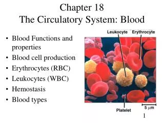
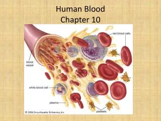
Composition of Whole Blood • Whole blood = plasma + formed elements • Plasma = matrix (fluid part of blood) • Plasma proteins in solution (vs. insoluble fibers like other CT) • Makes blood more dense than water • Formed elements = suspended blood cells/cell fragments
Composition of Whole Blood • Formed elements (formed through hematopoiesis) • RBCs (erythrocytes) • Most abundant • Transport O2 and CO2 • WBCs (leukocytes) • Part of immune system/defense mechanisms • Platelets • Small, non-cellular, membrane-bound packets of cytoplasm • Contain enzymes, substances important in clotting
Plasma • = H20 (> 90%) + plasma proteins + other solutes • Watery characteristic allows plasma to function as transportation medium for materials needed and no longer needed by body’s cells • Slightly more than half the blood volume • Plasma + interstitial fluid (IF) = most of the fluid outside cells (ECF, extracellular fluid) • Primary differences b/w plasma and IF • More dissolved protein in plasma (large size and globular shapes can’t cross capillary walls) • Higher levels of respiratory gases (O2, CO2) in IF due to activities of tissue cells
Classes of Plasma Proteins • Albumins (60%) • Globulins (35%) • Fibrinogen (4%) • Other (1%)
Albumins • Most abundant • Contribute to osmotic pressure • Transport • Fatty acids • Thyroid hormones • Steroid hormones
Globulins • Antibodies (immunoglobulins, IGs) • Attack foreign proteins and pathogens • Transport • Small metal ions (Fe) • Thyroid hormones • Other compounds that otherwise might be lost at the kidneys or have low solubility in water
Fibrinogen • Produces fibrin (long, insoluble, protein strands) • Forms blood clots • In a blood sample, if remove clot (with clotting proteins), remainder = serum • To prevent clot: use anticoagulant, e.g., EDTA, citrate • KNOW if sample is plasma or serum (for blood tests)
Other Plasma Proteins • Enzymes • Prohormones (proteins) • Hormones
Plasma Protein Synthesis • >90% by liver • Liver disease can lead to excess bleeding (inadequate fibrinogen and other clotting proteins) and other blood disorder • Plasma cells make antibodies • Endocrine organs make peptide hormones
Formed Elements • RBCs, WBCs, platelets • Formed in red bone marrow in adult (hematopoiesis) • Differentiation • Hemocytoblasts (stem cells) • Lymphoid stem cells lymphocytes (WBCs) • Myeloid stem cells RBCs, some WBCs, platelets • Erythropoiesis • RBC formation • Stimulated by erythropoietin (EPO) • Platelets formed from fragmentation of megakaryocytes
RBCs • Most abundant • 99.9% of formed elements and 1/3 of ALL body cells • Several million produced/sec • Most specialized blood cell • Contains hemoglobin that binds and transports O2 and CO2(primary function of both hemoglobin and RBCs) • Normal RBC count • Males: 4-6 million/uL Females: 4-5 million/uL • Androgens stimulate RBC production, estrogens do not
RBCs • Biconcave disc (thin in middle, thick at edge; like breath mint) • Large surface area to volume • Quickly absorbs and releases oxygen • Can form stacks (like breath mints) • Smooths flow through small blood vessels • Can bend and flex • To fit through small capillaries
RBCs • No nucleus, ribosomes, mitochondria • Cannot divide • Cannot synthesize enzymes or other proteins • Cannot perform repairs (lifespan = 120 days/3 mo) • Low energy demand • Gets energy from anaerobic metabolism of glucose absorbed from plasma • Transported O2 is NOT stolen by RBC for energy needs
RBCs • Hematocrit (packed cell volume, PCV) • % of formed elements in whole blood (most of which are RBCs) • Male = 46% • Female = 42% • Increases with dehydration, EPO • Decreases with internal bleeding, anemias, problems with RBC formation, e.g., sickle cell anemia
Hemoglobin (Hb) • Made of 4 globular protein subunits • 2 alpha chains; 2 beta chains • Each subunit contains 1 heme molecule • Organic ring structure around single Fe ion • Makes up most of RBC • Function • Allows cells to reversibly bind and transport O2 and CO2 on Fe • When tissue O2 low: O2 released from Hb, CO2 binds • When tissue O2 high: O2 binds to Hb, CO2 released
Diseases Involving Hemoglobin • Anemia • Inadequate tissue O2 levels O2 starvation • HCT too low • Hb content of RBCs reduced • Symptoms • Weakness, lethargy, mental confusion
Potentially Lethal Inherited Blood Disorders • From mutations that alter DNA sequence for hemoglobin • Thalassemia • Can’t produce enough alpha or beta chains of Hb • RBC production slowed, mature RBCs fragile and short-lived • Produces anemia • Sickle cell anemia • Change in amino acids of beta chain abnormal RBCs, lower O2 concentrations (because of abnormally shaped/defective Hb) • Cells can become stuck in capillaries circulatory block, cell death
Blood Types • Determined by antigens on surface of RBCs • Antigen = anything that can trigger an immune response; defense mechanism • >50 surface antigens on RBC, but A, B, and Rh are most important • 4 basic blood types • Blood type A = surface antigen A • Blood type B = surface antigen B • Blood type AB = surface antigens A and B (universal recipient) • Blood type O = neither A or B surface antigens (universal donor)
Blood Types • Plasma always contains antibodies that will react with foreign surface antigens, but not with “normal” • Type A blood: surface antigen A on RBCs and anti-B antibodies in plasma Fig. 19-7, p. 651
Blood Types • Rh positive = surface antigen Rh • Rh negative = no Rh surface antigen • Plasma anti-Rh antibodies present in Rh- ONLY if sensitized by previous exposure to Rh+ RBCs • Rh- mom has from first Rh+ baby Fig. 19-9, p. 654-655
Cross-reaction (Transfusion Reaction) • Same surface antigen and Ab agglutination (clumping) and hemolysis (cell death) • Plugs small vessels in vital organs: kidneys, lungs, heart, brain = fatal • Blood for transfusions must be carefully analyzed • Determine blood type: expose recipients blood to antibodies A and B • Cross-match: expose recipients blood to donor blood; reveals all cross-reactions Fig. 19-7b, p. 651
Today in class we will discuss: • White Blood Cells (WBCs) • Categories of WBCs based on their structure and function • The significance of changes in a differential count • Structure and Function of platelets • The definition of hemostasis • The mechanisms that control blood loss • The definition of hemopoiesis • The role of hemocytoblasts, lymphoid stem cells, myeloid stem cells, megakaryocytes, and reticulocytes • Locations of body sites used for blood collection and the basic physical characteristics of blood samples drawn from those sites
WBCs • WBCs vs. RBCs • Have nuclei and other organelles • Lack hemoglobin • 100s – 1000s of WBCs/uL vs. millions of RBCs/uL • Small fraction of WBCs circulate in blood • Lifespan = hrs (3 mo. for RBCs) • General functions • Defend body against invasion by pathogens and foreign proteins (lymphocytes) • Remove toxins, wastes • Attack abnormal or damaged cells (all other WBCs)
WBCs • Types of WBCs distinguished by staining techniques • Granulocytes – abundant (usually stained) granules (secretory vesicles and lysosomes) • Neutrophils - granules difficult to stain • Eosinophils - stain red-pink with acidic, red dye eosin • Basophils - stain deep purple-blue with basic dyes • Agranulocytes - have smaller stained granules; nuclei darkly stained • Monocytes • Lymphocytes
WBCs: Circulation • Small fraction of WBCs circulate in blood • Circulate for only small portion of life span (hrs) • Most in CT or lymphatic organs • Migrate through loose and dense connective tissue • Use blood stream: • To travel from one organ to another • For rapid transportation to areas of injury or invasion • When traveling through capillaries, can detect chemical signs of damage to surrounding tissues • Leave bloodstream to enter damaged area
WBCs: Movement • 4 characteristics of circulating WBCs • Amoeboid movements (like “the blob”) allows movement along walls of blood vessels and through tissues • Can move out of blood vessels (diapedesis) • Attracted to specific chemical stimuli (positive chemotaxis) • Guides WBCs to invading pathogens, damaged tissues, and active WBCs • Neutrophils, eosinophils, and monocytes capable of phagocytosis • Engulf pathogens, cell debris, and other materials • Neutrophils and eosinophils = microphages • Monocytes = macrophages
Hierarchy of WBCs • Lymphocytes (T cells, B cells, NKCs) • Phagocytes • Microphages • Eosinophils • Neutrophils • Macrophages • Monocytes • Basophils mast cells
WBCs • Neutrophils – 50-70% • Lymphocytes – 20-30% • Monocytes – 2-8% • Eosinophils – 2-4% • Basophils - < 1% • Mnemonic: Never Let Monkeys Eat Bananas Neutrophils Lymphocytes Monocytes Eosinophils Basophils
Neutrophils • Most abundant WBC in healthy individual (50-70% of circulating WBCs) • Nucleus has 2-5 lobes = polymorphonuclear leukocytes (PMNs) • Cytoplasm has pale granules • Contain lysosomal enzymes and bactericidal compounds • Highly mobile = FIRST WBC to arrive at injury site • Release hormones • Prostaglandins (coordinate local cellular activities) • Leukotrienes (coordinate tissue responses to injury or disease) • Phagocytic cells (microphages); specialize in attacking/digesting bacteria • 10-hr life span, only 30 min if actively engulfing
Figure 19-10a White Blood Cells RBC Neutrophil LM 1500
Lymphocytes • 20-30% of circulating WBCs • Smallest WBCs • Large nucleus with thin halo of cytoplasm • Most in CT and organs of lymphatic system • Part of the body’s specific defense system • e.g., T cells, B cells, natural killer cells (NKCs) • NKCs important in preventing cancer • Detect and destruct abnormal tissue cells
Figure 19-10e White Blood Cells RBC Lymphocyte LM 1500
Monocytes • 2-8% of WBCs • Large cells with large nucleus • Become macrophages • Very phagocytic • Engulf large particles and pathogens • Secrete substances that attract immune system cells and fibroblasts to injured area
Figure 19-10d White Blood Cells RBC LM 1500 Monocyte
Eosinophils • 2-4% of WBCs • Red/acid-staining granules • Two-lobed nucleus • Phagocytic cells (microphages) • Engulf bacteria, protozoa, cellular debris • Exocytose toxic compounds • Defend against large multicellular parasites (flukes, parasitic worms) • Also involved in allergic reactions
Figure 19-10b White Blood Cells RBC Eosinophil LM 1500
Basophils • <1% of circulating WBCs • Numerous granules; dark/stained with basic (blue-purple) dyes • Migrate to injury sites, release • Histamine • Dilates blood vessels • Heparin • Prevents blood clotting and promotes inflammation
Figure 19-10c White Blood Cells RBC Basophil LM 1500