Bacterial Pathogens
Bacterial Pathogens. John Scott Meschke Office: Suite 2338, 4225 Roosevelt Phone: 206-221-5470 Email: jmeschke@u.washington.edu. Turbidity. Pathogenic Bacteria.


Bacterial Pathogens
E N D
Presentation Transcript
Bacterial Pathogens John Scott Meschke Office: Suite 2338, 4225 Roosevelt Phone: 206-221-5470 Email: jmeschke@u.washington.edu
Pathogenic Bacteria Pathogenic bacteria possess virulence properties in the form of structures or chemical constituents that contribute to pathophysiology • Outer cell membrane of Gram negative bacteria: endotoxin (fever producer) • Exotoxins Pili: for attachment and effacement to cells and tissues Invasins: to invade cells Some bacteria make spores: • highly to physical and chemical agents and • very persistent in the environment Enteric and respiratory bacteria are important in environmental health
Peptidoglycan • Responsible for rigidity of the cell • 5-10% of wall mass in G- • 60-70% of wall mass in G+ • Not a permeability barrier • Site of action of penicillin- effects G+ much more than G- • Archebacteria have modified version
Outer envelope • Unique to G- bacteria • Can contain enzymes or enzyme like materials • Can act as permeability barrier but less so than plasma membrane • Contains endotoxins, antigens, a big mix of different materials
Capsule • Outside the cell wall- usually present only when cell has adequate carbon • Composed of polysaccharide- glycocalyx • External storage for carbon • Protect from phagocytosis • Glue
Endospores • Survival not reproduction • heat, drying, radiation, pH • Turned on by running out of nutrients • May be important in viable but not culturable phenomenon • Great economic cost • Major issue in food safety
Bacterial Taxonomy • Very little known about bacterial diversity, except in reference to illness • Early attempts at classifying bacteria were based morphological, biochemical, and serologic (phenotypic) properties • More recent classification methods also rely on genetic (and advanced phenotypic) methods • e.g. DNA-DNA hybridization studies, rRNA homology, etc. • Chemotaxonomy- FAA, WCPA, Cell wall composition, etc. • Polyphasic taxonomy approach results in fluidity at genus level and below
Actinobacteria Aquificae Bacteroidetes Chlamydiae Chlorobi Cyanobacteria Deinococcus Firmicutes Fusobacteria Proteobacteria Spirochaetes Thermotogae Bacterial Phyla
Actinobacteria Corynebacteria Mycobacteria Bifidobacteria Bacteroidetes Porphyromonas Chlamydiae Chlamydia Fimicutes Bacilliales Bacillus Listeria Staphylococcus Clostridiales Clostridia Lactobacillales Enterococcus Lactobacillus Lactococcus Streptococcus Mollicutes Mycoplasma Important Genera
Proteobacteria Alphaproteobacteria Bartonella Brucella Rickettsia Betaproteobacteria Bordatella Burkholderia Neisseria Epsilonproteobacteria Campylobacter Helicobacter Important Genera
Proteobacteria Gammaproteobacteria Aeromonadaceae Aeromonas Enterobacteriales Escherichia Salmonella Shigella Yersinia Legionellales Coxiella Legionella Gammaproteobacteria Pasturellales Pasturella Haemophilus Pseudomonadales Pseudomonas Thiotrichales Francisella Vibrionaceae Vibrio Important Genera
Spirochaetes Leptospira Borrelia Treponema Important Genera
Bacillus anthracis: Anthrax • Large, gram positive non-motile rod • Vegetative form and spores • Nearly worldwide distribution • Over 1,200 strains • 5th Plaque of Bible • Spores 1.5-3 microns
Epidemiology • Three forms of natural disease • Inhalational • Rare (<5%) • Most likely encountered in bioterrorism event • 86-100% Mortality (despite treatment) • Cutaneous • Most common (95%) • Direct contact of spores on skin • <5% (treated) – 20% (untreated) mortality • Gastrointestinal • Rare (<5%), never reported in U.S. • Ingestion • approaches 100% mortality
Inhalational Anthrax • Infective dose = 8,000 - 15,000 spores • Incubation period = 1-6 days • Duration of illness = 3-5 days • Fever, malaise, and fatigue • Short period of improvement = up to 2 days • Abrupt respiratory distress…death <24hrs • No person to person transmission
Anthrax: Cutaneous • Most common form (95%) • Inoculation of spores under skin • Incubation: hours to 7 days • Small papule ulcer surrounded by vesicles (24-28h) • Painless eschar with edema • Death 20% untreated; rare if treated USAMRICD: Eschar with surrounding edema
Anthrax: Gastrointestinal • Ingestion of contaminated meat • Incubation: hours or up to 7 days • Fever, acute gastroenteritis, vomiting, bloody diarrhea
Stapylococcus • A leading cause of bacteremia • Virulence leads to a high proportion of endocarditis (including on normal heart valves), metastatic infections, and/or death • Staphylococcus aureus bacteremia is both a cause and a result of endocarditis • Changing epidemiology is a therapeutic challenge • Growing resistance to beta-lactam antibiotics • Increasing tolerance to vancomycin
Staphylococcus aureus • Gram positive, aerobic cocci • Staph are found in air, dust, sewage, water milk, food, equipment, animals and humans: skin, hair, nose, throat, open sores, boils, saliva • Transmitted to foods via handling, coughing, sneezing, wiping
Clostridium perfringens • Gram positive, spore forming, anaerobic rod • Found in soil, intestinal tracts of man and animals • Foodborne infection;8-22 hours; toxin is formed in the gut • Symptoms: Diarrhea, severe dehydration, cramps
Clostridium perfringens • Large number of cells (108) needed to cause disease • Associated foods: temperature abused foods, roast beef, stews, meat gravy, poultry
Campylobacters • Gram-negative • Curved rod • about 1.5-3 microns • motile via polar flagella • Microaerophilic • Prefer high CO2
Campylobacters • Due to Campylobacter jejuni or sometimes C. coli. • Normal intestinal flora of many warm-blooded animals • chickens and turkeys; also in raw water and raw milk. • Illness in sheep (abortion), dogs and cats (gastroenteritis) • Causes illness in humans • Cause 5-11% of all diarrhea cases in the United States. • Symptoms: from mild to severe: • bloody diarrhoea is the most characteristic symptom) • also fever, nausea, abdominal cramps and (seldom) vomiting • duration of illness usually 2-10 days • abdominal cramps, may recur for up to 3 months after infection • Complications such as septicaemia, may arise. • The infectious dose may be very low, i.e. 100s of cells • Infants, young children and debilitated people at highest risk
Complications and Sequelae of Campylobacteriosis; Guillain-Barre Syndrome • Develop a rare disease of the nervous system beginning several weeks after the diarrheal illness. • Called Guillain-Barré syndrome • Person's immune system is "triggered" to attack the body's own nerves • can lead to paralysis lasting several weeks; usually requires intensive care • About 1 per 1000 reported campylobacteriosis cases leads to Guillain-Barré syndrome. • Perhaps 40% of Guillain-Barré syndrome cases in this country may be triggered by campylobacteriosis.
Escherichia coli cells: ~0.5 x 1.0 micrometers Typical rod-shaped bacteria: fecal indicator and pathogenic strains
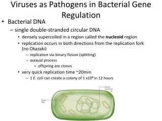
E. coli Genetics and Serology Genetics: • Single, circular DNA molecule, ~4 x 10⁶ base pairs • Molecular weight of 4 x 10⁹ • Total length of about 1.4mm. • Two strains completely sequenced and genomic organization is now being characterized • many of the genes have been mapped. Serology: • E. coli can be subdivided by somatic (cell-wall) or O antigens and flagellar or H antigens. • >160 recognized O types and 55 recognized H types • over 8000 possible OH serotypes. • also capsular (K) and fimbrial antigens.
Virulence Properties of E. coli Enterotoxins: • at least two types: Heat Stable (ST) and Heat Labile (LT) • Verotoxins or Shiga-like toxins (interchangeable terms): • Verotoxin term is based on the reactions of toxins on Vero cells • at least two families of these toxins: • VT1 (SLT I): similar to Siga-toxin (produced by some strains of Shigella dysenteriae) • VT2 (SLT II) which is only about 50% realted Shiga toxin. • Other Toxins: • Cytolethal distending toxin (CLDT), VirCytotoxin, Cytotoxic necrotising factors (CNF), a possible Enteropathogenic E. coli EPEC) enterotoxin and a possible E. coli Sudden Infant Death Syndrome (SIDS)-toxin. • Haemolysins: • extracellular haemolysin known as alpha-haemolysin (many strains) • cell-associated haemolysin, beta-haemolysin, (some strains) • enterohaemolysin: extracellular; Enterohaemorrhagic E. coli (EHEC)
Virulence Properties of E. coli • Fimbriae: CFAI/CFAII, Type 1 fimbriae, P fimbriae, S fimbriae • most important: K88, K99 and CFA fimbriae associated with enterotoxigenic E. coli (ETEC). They have differing species specificities. • The p-fimbriae: associated with urinary tract pathogens. • E. coli also produce common fimbriae not associated with virulence. • Adhesins: • Intimin: non-fimbrial adhesin; causes the intimate association with target cells in enteropathogenic and enterohaemorrhagic E. coli . • Associated with the 'attachment and effacement' phenomenon • Causes destruction of the intestinal surface cells. • Other outer membrane proteins can act as adhesins.
Pathogenic E. coli Enteric Infections: • Enteroadherent E. coli (EAEC) • Enteroaggregative E. coli (EAggEC) • Enterohaemorrhagic E. coli (EHEC) • Enteroinvasive E.coli (EIEC) • Enteropathogenic E. coli (EPEC) • Enterotoxigenic E. coli (ETEC) Extraintestinal Infections: • Uropathogenic E. coli (UPEC): urinary tract infections • Neonatal Menigitis E. coli (NMEC).
Enterohemorrhagic E. coli Harbor genes for one or more of the virulence attributes known to associated with the EHEC. • shiga toxin(s) • adherence factor(s) • enterohaemolysin • somatic antigens characteristic of many EHEC serogroups, such as O111 or O157. • An E. coli must cell carry a sufficient number of such genes to cause disease. • Magnitude of exposure or size of infectious dose is also important. • Dose is very small in comparison with those for most other enteric pathogens; a few bacteria per dose may cause infection and illness
E. coli O157:H7 • As few as 10 cells may cause disease • Associated foods: undercooked hamburgers, venison jerky, water, unpasteurized milk, fermented sausages, sprouts, water, roast beef, apple cider, salami, lettuce, yogurt, cantaloupe
Enteropathogenic E. coli • Cause infantile gastroenteritis. • Certain serotypes are associated with infantile diarrhea. • Infantile gastroenteritis with dehydration is an important problem. • Serogroups O26, O55, O111, O119, O125, O126, O127, and O128 are most commonly isolated. • EPEC have declined in the developed world as major causes of infantile diarrhea, but remaining very important in the developing world. • EPEC adhere to the intestinal mucosa to produce a characteristic "attaching and effacing" lesion in the brush border microvillous membrane. • EPEC trains belonging to serogroups O26, O111 and O128 have recently emerged as enterohaemorrhagic E. coli (EHEC).
Enterotoxigenic E. coli • Major cause of travellers' diarrhea (Montezuma’s revenge, Delhi belly, Aztec two-step, etc.) and of diarrhea in children in the developing world • Produce an enterotoxin similar to cholera toxin. • They are involved with a condition known as "Non-Vibrio cholerae cholera-like diarrhoea". • Produce one or both of two enterotoxins: • heat stable enterotoxin (ST) : survives boiling for 30 minures • heat labile enterotoxin (LT): does not survive such boiling • LT response is sensitive to acid pH; ST was not. • LT is closely related to choleragen (CT) the enterotoxin of V. cholerae. • Most ETEC isolated from humans produce colonization factor antigens which are human specific fimbrial antigens • also a common cause of diarrhea in young animals.
Enteroinvasive E. coli (EIEC) • Cause positive reaction in the Sereny test (ability to cause keratoconjunctivitis in guinea pig eyes). • a characteristic shared with strains of Shigella • By DNA probes the invasiveness plasmids of both E. coli and Shigella are identical. • 120-140 mD invasiveness plasmids encode all the genes necessary for the virulence of the EIEC. • Many EHEC are non-motile and anaerogenic. • Account for only a small proportion of diarrhea in non-tropical countries • but, cause high proportions of illness and death, mostly in warmer seasons • Also important causes of dysentery-like diarrhea in tropical countries.
Enteroadherent E. coli (EAEC) • Different patterns of adherence to cultured epithelial cells. • Localized adherent E. coli (LAEC) by some serotypes • form adherent microcolonies on HEp-2 cells • associated with acute, non-bloody diarrhoea in children. • Diffusely adhering E. coli (DAEC) • a cause of diarrhea in some studies
Enteroaggregative E. coli (EAggEC) • 3rd type of adherence: enteroaggregative adherence • Bacteria align in parallel rows to cells or glass ('stacked brick-like’). • persistent childhood diarrhoea in South America and India • Illness lasts >14 days. • Produce a heat-labile toxin antigenically related hemolysin but not hemolytic • Produce plasmid encoded heat stable toxin (EAST1) unrelated to the heat stable enterotoxin of ETEC. • Form a distinct “scum” on the surface of Mueller-Hinton broth • possibly use as a rapid screening method for EAggEC.
Uropathogenic E. coli (UPEC) • Common cause of urinary tract infections (UTI). • Severity: from asymtomatic to bacteriuria, cystitis and pyelonephritis. • Women more frequently affected than men. • Same serotypes are found in feces and urine of patients, but, UPEC have virulence factors which enhance their ability to cause infection. • Only some O groups cause UTI: O1, O2, O4, O6, O7, O18 and O75. • Only some K antigens (K1, K2, K3, K5, K12 and K13) cause UPEC. • Certain pili (fimbrae), the p-pili, are an important virulence factor • bind to the P-antigen, a blood grouping antigen • bind more to uroepithelial cells of persons with the P or P2 phenotype • other adhesins may also be involved. • Production of alpha-haemolysin by UPEC • Production of aerobactin • Several virulence enhance an E. coli's ability to cause UTI • an infecting strain may only express some of them to cause infection.
Neonatal Meningitis E. coli (NMEC) • Neonatal menigitis in about 1/2500 live births. • Up to 80% of cases of neonatal menigitis are due to E. coli. • ~80% of the isolates possess the K1 capsular antigen. • a 2.8 a-linked homopolymer of N-acetylneuraminic acid (sialic acid) • chemically and immunologically identical to the group B acidic polysaccharide of Neisseria menigitidis. • masks the underlying structures of the bacterial cell surface, preventing specific antibody responses and the activation of the alternate complement system from being activated.\ • poor immunogen, maybe due to it resembling extracellular matrix proteins. • may also reduce the serum sensitivity of E. coli. • serotypes isolated from meningitis cases often found in maternal faeces; source; infection occurs at birth. • Reasons for susceptibility of neonates to NMEC is not known.