The Cardiovascular System
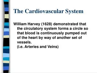
The Cardiovascular System. William Harvey (1628) demonstrated that the circulatory system forms a circle so that blood is continuously pumped out of the heart by way of another set of vessels. (i.e. Arteries and Veins). The Cardiovascular System.

The Cardiovascular System
E N D
Presentation Transcript
The Cardiovascular System William Harvey (1628) demonstrated that the circulatory system forms a circle so that blood is continuously pumped out of the heart by way of another set of vessels. (i.e. Arteries and Veins)
The Cardiovascular System Heart attacks are, perhaps, the number one killer in the USA. In middle age and older persons, the probability of developing a coronary disease increases with: 1. Smoking 2. Obesity 3. Cholesterol/triglycerides 4. Abnormal ECG 5. Hypertension
I. Blood Vascular A. Heart B. Arteries C. Capillaries D. Veins II. Lymphatic Vascular A. Lymphatic Capillaries B. Lymphatic Vessels Thoracic Duct Right Lymphatic Duct The Cardiovascular System
Impulse Conduction A-V Node Junctional fibers (0.01m/s) Node (0.1m/s) Transitional (0.4m/sec) Atrionodal Nodal Nodal His Bundle of His Branches of bundle of His E Purkinje fibers rate L5 to 4.0 m/sec
Cardiac Action Potentials Pace-maker -90 -60 40 Atrial Myocradial cell 2 1. Resting potential 2. Spike (rapid depolarization) 3. Plateau (Ca++ perinmeability ) 4. Repolarization 3 No+ in 4 Co in K+ set 1 -90 Ventricular Myocradial cell
I. Unipolar (V) leads (9 locations) A. Chest leads (precordial) V 1- 6 B. Limb leads VR (right arm) VL (left arm) VF (left foot) II. Bipolar Limb Leads I. from RA to LA II. from RA to LL III. from LA to LL R T P Q S Electrocardiogram (ECG) The ECG is the algebraic sum of the electrical events which occur during a heart beat. These signals are transmitted via the body fluids to the body surface where they are recorded by electric amplifiers. The series of pikes and depressions have arbitrarily been labeled
Relationship between Monophasic Action Potential and Ventricular Activity Depolarization Repolarization R T Q S
R T P Q S ECG PR interval ST segment Duration Event Ave. Range PR interval 0.18 0.12-0.20 Atrial Dep. Through AV QRS duration 0.08 0.006-0.10 Aent. Dep. QT interval 0.40 0.37-0.43 Vent. Dep + Repol. ST interval 0.32 Vent. Repolarization PR segment QT interval QRS Duration
Pathological Conditions Mean Electrical Axis (mark changes in current flow) Left Axis Deviation (hypertrophy of left ventricle) 1. Hypertension (muscle mass on left side of heart) 2. Aortic Valvular stenosis 3. Aortic Valvular regurgitation 4. Several congenital heart conditions Right Axis Deviation 1. Hypertrophy of right ventricle (pulmonary stenosis) 2. Tetralogy of Fallot (congenital right to left shunt )
Cardiac Arrhythmias I. Correlation of Plasma K+ Level a. Noraml (plasma K+ 4 - 5.5 mEq/l) b. Hypokalemia (plasma K++ 2.5 mEq/l) c. Hypokalemia (plasma K++ 2.3 mEq/l) d. Hyperkalemia (K++ 7.0) e. Hyperkalemia (K++ 8.5 II. Atrial Enlargement a. “p” pulmonale b. “p” mitrale u p t u t
Origin of the heartbeat & the electrical activity of the heart
Cardiovascular Pressures & Sounds aortic valve opens closes Dicrotic notch 120 - 100 - 80 - 60 - 40 - 20 - 0 - Key (color) 1. Left ventricular P. 2. Aortic pressure 3. Left atrial p. a v c closes opens mitral valve Electrocardiogram Phonocardiogram 1. Lub 2. Dub 3. Rapid ventricular filling 4. Atrial contraction 4 1 2 3
Comparison of Pressures Within the Circulatory System Aorta Venale Arteriale capilleries Large veins Terminal veins Terminal arteries Large arteries Vena cava Main venous branch Main arterial branch 120 100 80 60 40 20 0 Systolic Pressure Diastolic Pressure
Time seconds Heart Sounds 0 0.2 0.4 0.6 0.8 120- 100- 80- 60- 40- 20- 0 Ventricle Pressure (mmHg) Disstole Systole 1 120- 80- 40- 6 5 Volume (ml) 2 3 4 1st 2nd 3rd Heart sounds
Heart Murmurs(Bruits - Outside the heart) Types: A Innocent (functional) B pathogenics (non-functional) 1. Stenosis - narrowing of valve 2. Regurgitant (insufficient) 3. Prolaspse C. Congential 2. Patent ductus arteriosis 2. Interventricular septal defect
Aortic or Pulmonary Mitral or Tricuspid Heart Murmurs(Bruits - Outside the heart) Valve Name Abnormality Timing of Murmur Stenosis Systolic Regurgitation Diastolic Stenosis Diastolic Regurgitation Systolic Rheumatic Vascular lesions cause an autoimmune disease in which the heart valves are likely to be damaged . It is caused by a group A streptococcal toxin such as the ones which cause sore throat, scarlet fever, or middle ear infection.
Heart & Circulation 1.Blood Blood cells become packed at the bottom of the tube when whole blood is centrifuged, leaving the fluid at the top of the tube. Red blood cells are the most abundant of the blood cells -- white cells & platelets float ?? Only a thin, light-colored “buffy coat” at the interface between the packed red blood cells and the plasma.
Representative normal plasma values Measurement Normal Range Blood volume 80-85 ml/kg body weight Blood osmoiality 280-296 mOsm Blood pH 7.35-7.45 Enzymes: Creatine Female: 10-79 U/L phosphokinase (CPK) Male : 17-148 U/L Lactic Dehydrogenase 45-90 U/L (LDH) Phosphate (acid) Female: 0.01-0.56 Sigma U/ml Male: 0.13-0.63 Sigma
Representative normal plasma values Measurement Normal Range Hematology Values: Hematocrit Female: 37%-48% Male: 45%-52% Hemoglobin Female: 12-15 g/100ml Male: 13-18 g/100ml Red blood cell count 4.2-5.9 million/mm3 White blood cell count 4,300-10,880/mm3 Hormones Testosterone Male: 300-1,100ng/100ml Adrenocorticotropic (ACTH) 15-70 pg/ml Growth hormone Children: over 10ag/ml Adult male: below 5 mg/ml Insulin 6-26 * U/ml (fasting)
Representative normal plasma values Measurement Normal Range Ions Bicarbonate 24-30mmol/l Calcium 2.1-2. 6mmol/l Chloride 100-106mmol/lPotassium 3.5-5.0mmol/l Sodium 135-145mmol/1
Representative normal plasma values Measurement Normal Range Organic Molecules (other) Cholesterol 120-220mg/100ml Glucose 70-110mg/100ml (fasting) Lactic acid 0.6-1.6mmol/l Protein (total) 6.0-8.4g/100ml Triglyceride 40-150mg/100ml Urea nitrogen 8-25mg/100ml Uric acid 3-7 mg/100ml
Hemostasis Mechanisms 1. Vascular Spasms (vasoconstricitions) 2. Formation of Platelet Plug 3. Blood Coagulation
Table 13.4 Plasma Clotting Factors * * + + * * + * Require Vitamin K + Antihemophilic factors
Table 13.5 Some acquired and inherited defects in the clotting mechanism
Common pathway Intrinsic pathway Extrinsic pathway
Extrinsic pathway Activator: tissue thromboplastin VII VII activated VII complex (VII, tissue thromboplastin, calcium, phospholipids) Common pathway
XII XII activated XI XI activated IX IX activated Intrinsic pathway Activators: collagen,gas, and others VIII complex (VIII, (X activated, calcium, phospholipids) Common pathway
X X activated V complex (V, X activated, calcium, phospolipids Prothrombin Thrombin Fibrinogen Fibrin Extrinsic pathway Common pathway Intrinsic pathway Fibrin polymer XIII
Prevention of Blood Clotting A. In-Vivo 1. Endothelial Surface Factors (Prostocyclin) 2. Heparin: A conjugated polysaccharide produced by mast cells (basophils) 3. Coumarin(Warfarin) & Phenindione: orally administered; blocks the synthesis of Vit.-K dependent clotting factors 4. Aspirin
Prevention of Blood Clotting B. In-Vitro 1. Removal of Ca++ a. Oxalates b. Citrates c. Chelating agents (EDTA) 2. Inhibition of vitamin-K Dicumarol (coumarin) • 3. Aspirin Inhibits platelet aggregation
O C O O CH2 C CH2 O N Me 2+ CH2 O CH2 N C CH2 O O CH2 C O EDTA A chelate of ethylenediamine tetraacetate with a divalent metal cation (Me2+). The shaded portion represents the plane of the coordination bonds
Causes for Excessive Bleeding 1. Vitamin-K Deficiency Factors II, VII, IX & X 2. Hemophilia Factors VII (75%), IX (15%), & XI (5-10%) 3. Thrombocyopenia
Vessel Damage Exposed Collagen Extrinsic Pathway Intrinsic Pathway Vessel Damage Subendothelial Cells Exposed to Blood Contact Activation Tissue Factor XII XIIa VIIa XIa XI VII Ixa IX IX VIII Activated Platelets XIIIa Xa X X Activated Platelets Va V Prothrobin Thrombin