BEA and ENN Cyclohexadepsipeptide Toxicity Effects
Study on the ionophoric properties of BEA and ENN cyclohexadepsipeptides, exploring their impact on cellular metabolic state and ionic homeostasis.

BEA and ENN Cyclohexadepsipeptide Toxicity Effects
E N D
Presentation Transcript
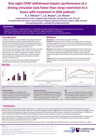
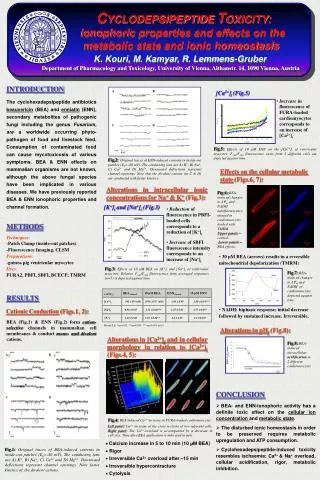
(-ΔF/Fo) BEAControl 10 μM ENN ENNControl 10 μM BEA CYCLODEPSIPEPTIDE TOXICITY: ionophoric properties and effects on the metabolic state and ionic homeostasisK. Kouri, M. Kamyar, R. Lemmens-GruberDepartment of Pharmacology and Toxicology, University of Vienna, Althanstr. 14, 1090 Vienna, Austria [Ca2+]i 542 50 (nM) 2656 55* (nM) 2.80 0.24*** 1.68 0.09 [Na+]i 0.98 0.07 1.47 0.01 1.31 0.08*** 1.75 0.03* [K+]i 1.49 0.06 2.2 0.10* 2.6 0.05 1.21 0.08** 6 0 3 12 8 10 13 14 15 [Ca2+]i (Fig.5) Increase in fluorescence of FURA-loaded cardiomyocytes corresponds to an increase of [Ca2+]i [K+]i and [Na+]i (Fig.3) Reduction of fluorescence in PBFI-loaded cells corresponds to a reduction of [K+]i Increase of SBFI fluorescence intensity corresponds to an increase of [Na+]i 0 6 Fig.5:Effects of 10 μM ENN on the [Ca2+]i of ventricular myocytes. F340/F380 fluorescence ratio from 5 different cells are depicted against time. Fig.2:Original traces of ENN-induced currents in inside-out patches (Eh=-40 mV). The conducting ions are A) K+, B) Na+, C) Ca2+ and D) Mg2+. Downward deflections represent channel openings. Note that the divalent cations (in C & D) are conducted with faster kinetics. Effects on the cellular metabolic state (Figs.6, 7): Fig.3:Effects of 10 μM BEA on [K+]i and [Na+]i of ventricular myocytes. Relative F340/F380 fluorescence from averaged responses (n=5) is depicted against time. 2 pA 20 ms Alterations in intracellular ionic concentrations for Na+ & K+ (Fig.3): Fig.6:BEA-induced changes in ΔΨm and NADH autofluorescence imaged in cardiomyocytes loaded with TMRM.Upper panels = control.Lower panels = BEA effects. MeanS.E. *=p<0.05, **=p<0.005, ***=p<0.001 (n=5) 30 μM BEA (arrows) results in a reversible mitochondrial depolarization (TMRM) Fig.7:BEA-induced changes in ΔΨm and NADH in cardiomyocytes depicted against time. NADH: biphasic response; initial decrease followed by sustained increase. Irreversible. Alterations in pHi (Fig.8): Alterations in [Ca2+]i and in cellular morphology in relation to [Ca2+]i (Figs.4, 5): Fig.8:BEA-induced intracellular acidification in 2 different cardiomyocytes. A B A B Fig.4: BEA induced Ca2+ increase in FURA-loaded cardiomyocytes Left panel: Ca2+ increase at the cross sections of two adjacent cells. Right panel: The Ca2+-overload is accompanied by a decrease in cell-size. Time after BEA application is indicated in min. C D C D • Calcium increase in 5 to 10 min (10 μM BEA) • Rigor • Irreversible Ca2+ overload after 15 min • Irreversible hypercontracture • Cytolysis INTRODUCTION The cyclohexadepsipeptide antibiotics beauvericin (BEA) and enniatin (ENN), secondary metabolites of pathogenic fungi including the genus Fusarium, are aworldwide occurring phyto-pathogen of food and livestock feed. Consumption of contaminated food can cause mycotoxicosis of various symptoms. BEA & ENN effects on mammalian organisms are not known, although the above fungal species have been implicated in various diseases. We have previously reported BEA & ENN ionophoric properties and channel formation. METHODS Techniques: -Patch Clamp (inside-out patches) -Fluorescence Imaging, CLSM Preparations: -guinea pig ventricular myocytes Dyes: FURA2,PBFI, SBFI, BCECF; TMRM RESULTS Cationic Conduction(Figs.1, 2): BEA(Fig.1) & ENN (Fig.2) form cation-selective channels in mammalian cell membranes & conduct mono- and divalent cations. CONCLUSION • BEA- and ENN-ionophoric activity has a definite toxic effect on the cellular ion concentration and metabolic state. • The disturbed ionic homeostasis in order to be preserved requires metabolic upregulation and ATP consumption. • Cyclohexadepsipeptide-induced toxicity resembles ischaemia: Ca2+ & Na+ overload, cellular acidification, rigor, metabolic inhibition. Fig.1:Original traces of BEA-induced currents in inside-out patches (Eh=-40 mV). The conducting ions are A) K+, B) Na+, C) Ca2+ and D) Mg2+. Downward deflections represent channel openings. Note faster kinetics of the divalent cations.