Protein Structure Prediction
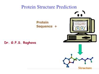
Protein Structure Prediction. Protein Sequence +. Dr. G.P.S. Raghava. Structure. Protein Structure Prediction. Experimental Techniques X-ray Crystallography NMR Limitations of Current Experimental Techniques Protein DataBank (PDB) -> 23000 protein structures

Protein Structure Prediction
E N D
Presentation Transcript
Protein Structure Prediction Protein Sequence + Dr. G.P.S. Raghava Structure
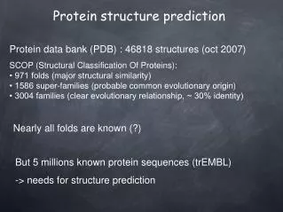
Protein Structure Prediction • Experimental Techniques • X-ray Crystallography • NMR • Limitations of Current Experimental Techniques • Protein DataBank (PDB) -> 23000 protein structures • SwissProt -> 100,000 proteins • Non-Redudant (NR) -> 10,00,000 proteins • Importance of Structure Prediction • Fill gap between known sequence and structures • Protein Engg. To alter function of a protein • Rational Drug Design
Protein Architecture • Proteins consist of amino acids linked by peptide bonds • Each amino acid consists of: • a central carbon atom • an amino group • a carboxyl group and • a side chain • Differences in side chains distinguish the various amino acids
Amino Acid Side Chains Vary in: • Size • Shape • Polarity
Conformation Flexibility • Backbone (main chain of atoms in peptide bonds, minus side chains) conformation: • Torsion or rotation angles around: • C-N bond () • C-C bond () • Sterical hinderance: • Most – Pro • Least - Gly
Protein Secondary Structure Secondary Structure Regular Secondary Structure (-helices, -sheets) Irregular Secondary Structure (Tight turns, Random coils, bulges)
Secondary Structure:Helices ALPHA HELIX : a result of H-bonding between every fourth peptide bond (via amino and carbonyl groups) along the length of the polypeptide chain Individual Amino acid H-bond
Helix formation is local THYROID hormone receptor (2nll)
Secondary Structure:Beta Sheets BETA PLEATED SHEET: a result of H-bonding between polypeptide chains
Definition of -turn A -turn is defined by four consecutive residues i, i+1, i+2 and i+3 that do not form a helix and have a C(i)-C(i+3) distance less than 7Å and the turn lead to reversal in the protein chain. (Richardson, 1981). The conformation of -turn is defined in terms of and of two central residues, i+1 and i+2 and can be classified into different types on the basis of and . i+1 i+2 i H-bond i+3 D <7Å
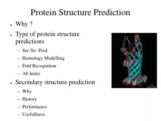
Tertiary Structure: Hexokinase (6000 atoms, 48 kD, 457 amino acids) polypeptides with a tertiary level of structure are usually referred to as globular proteins, since their shape is irregular and globular in form
What determines fold? • Anfinsen’s experiments in 1957 demonstrated that proteins can fold spontaneously into their native conformations under physiological conditions. This implies that primary structure does indeed determine folding or 3-D stucture. • Some exceptions exist • Chaperone proteins assist folding • Abnormally folded Prion proteins can catalyze misfolding of normal prion proteins that then aggregate
Levels of Description of Structural Complexity • Primary Structure (AA sequence) • Secondary Structure • Spatial arrangement of a polypeptide’s backbone atoms without regard to side-chain conformations • , , coil, turns (Venkatachalam, 1968) • Super-Secondary Structure • , , /, + (Rao and Rassman, 1973) • Tertiary Structure • 3-D structure of an entire polypeptide • Quarternary Structure • Spatial arrangement of subunits (2 or more polypeptide chains)
Techniques of Structure Prediction • Computer simulation based on energy calculation • Based on physio-chemical principles • Thermodynamic equilibrium with a minimum free energy • Global minimum free energy of protein surface • Knowledge Based approaches • Homology Based Approach • Threading Protein Sequence • Hierarchical Methods
Energy Minimization Techniques Energy Minimization based methods in their pure form, make no priori assumptions and attempt to locate global minma. • Static Minimization Methods • Classical many potential-potential can be construted • Assume that atoms in protein is in static form • Problems(large number of variables & minima and validity of potentials) • Dynamical Minimization Methods • Motions of atoms also considered • Monte Carlo simulation (stochastics in nature, time is not cosider) • Molecular Dynamics (time, quantum mechanical, classical equ.) • Limitations • large number of degree of freedom,CPU power not adequate • Interaction potential is not good enough to model
Molecular Dynamics • Provides a way to observe the motion of large molecules such as proteins at the atomic level – dynamic simulation • Newton’s second law applied to molecules • Potential energy function • Molecular coordinates • Force on all atoms can be calculated, given this function • Trajectory of motion of molecule can be determined
Knowledge Based Approaches • Homology Modelling • Need homologues of known protein structure • Backbone modelling • Side chain modelling • Fail in absence of homology • Threading Based Methods • New way of fold recognition • Sequence is tried to fit in known structures • Motif recognition • Loop & Side chain modelling • Fail in absence of known example
Homology Modeling • Simplest, reliable approach • Basis: proteins with similar sequences tend to fold into similar structures • Has been observed that even proteins with 25% sequence identity fold into similar structures • Does not work for remote homologs (< 25% pairwise identity)
Homology Modeling • Given: • A query sequence Q • A database of known protein structures • Find protein P such that P has high sequence similarity to Q • Return P’s structure as an approximation to Q’s structure
Threading • Given: • sequence of protein P with unknown structure • Database of known folds • Find: • Most plausible fold for P • Evaluate quality of such arrangement • Places the residues of unknown P along the backbone of a known structure and determines stability of side chains in that arrangement
Hierarcial Methods Intermidiate structures are predicted, instead of predicting tertiary structure of protein from amino acids sequence • Prediction of backbone structure • Secondary structure (helix, sheet,coil) • Beta Turn Prediction • Super-secondary structure • Tertiary structure prediction • Limitation Accuracy is only 75-80 % Only three state prediction