Chapter 19
Chapter 19. The Reproductive System. Introduction. Male reproductive system Anatomy: testes, scrotum, spermatozoa, semen, penis Physiology: ducts of the system; testosterone Female reproductive system Anatomy: ovaries, fallopian tubes, uterus, vagina, external genitalia, mammary glands

Chapter 19
E N D
Presentation Transcript
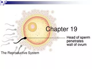
Chapter 19 The Reproductive System
Introduction • Male reproductive system • Anatomy: testes, scrotum, spermatozoa, semen, penis • Physiology: ducts of the system; testosterone • Female reproductive system • Anatomy: ovaries, fallopian tubes, uterus, vagina, external genitalia, mammary glands • Physiology: menstrual cycle, estrogen • Pregnancy, embryonic development
Male Reproductive System • Primary sex organs: testes (male gonads, aka testicles) • Produce sperm as exocrine glands; hormones as endocrine glands • Accessory structures
Testes • Paired oval glands approx 2” in length, 1” in diameter • Each testis is divided into lobules, containing tightly coiled seminiferous tubules, which produce sperm by spermatogenesis • Contained within scrotum
Scrotum • Outpouching of the abdominal wall • Loose skin and superficial fascia • Supporting structure of the testes • Internally separated by a septum into two lateral portions • Cremaster muscle located in spermatic cord contracts scrotal sac, and elevates testes • Maintains temperature about 3° below body temp, for ideal sperm and hormone production
Spermatozoa • Mature sperm cells are produced at a rate of 300 million/day • Life expectancy once ejaculated is ~ 48 hrs • 3 parts • Head, contains genetic material, acrosome • Midpiece, contains mitochondria • Tail, flagellum, locomotion
Testosterone • Controls development, growth, and maintenance of male sex organs • Causes descent of testes from abdomen • During puberty, stimulates bone growth • Stimulates protein build up in muscles • Stimulates maturation of sperm cells • Causes enlargement of thyroid cartilage (Adam’s apple), deepening of voice • Aggression, hair patterns and recession
Ducts of the System • Sperm cells are moves from the convoluted seminiferous tubules into straight tubules, then eventually empty into the ductus epididymis • Epididymis: comma-shaped structure on posterior of testis, where sperm cells mature • Sperm continues to move from epididymis into vas (ductus) deferens, then upward through spermatic cord into the abdomen
Pathway of sperm • Sperms travels through the vas deferens, where it joins with ducts from the prostate, and the urethra, becoming the ejaculatory duct • Secretions from the bulbourethral glands join the fluid as it becomes seminal fluid (semen), which is expelled through the urethra during ejaculation.
Semen • A mixture of sperm cells, and secretion from the prostate, bulbourethral glands, and seminal vesicles. • Milky in color, and sticky – fructose that provides energy for sperm • Alkaline, pH of 7.2 – 7.6 • Average volume 2.5 – 6 mL / ejaculation
Penis • Shaft • Glans penis – distal head • Prepuse – foreskin, removed during circumcision • Internally, spongy sinuses engorge with blood during erection
Female Reproductive System • Primary sex organs – ovaries (female gonads), paired • Produce ova (ovum), eggs as exocrine glands • Produce estrogen and progesterone as endocrine glands • Accessory structures: fallopian tubes, uterus, vagina, external genitalia
Ovaries • Located in upper pelvic cavity • Size of unshelled almonds • Held in position by suspensory ligaments
Ovum • Each ovary contains ovarian follicles in various stages of development • Oocyte is an immature follicle • Mature egg is called a graafian follicle • After egg ruptures from follicle, it changes into the corpus luteum (“yellow body”) which produces estrogen and progesterone
Fallopian Tubes • Paired tubes, transporting ova from each ovary to the uterus • Infundibulum is a funnel-shaped open end; lies close to, but not attached to, ovary • Fimbriae surround infundibulum and ovary
Uterus • In pelvic cavity • Between rectum and bladder • Where fertilized egg implants • Narrow opening is cervix • Fundus at top • Endometrium
Cervix • Opens into vagina • Isthmus small constricted region • Interior is cervical canal • Internal os opens into uterine cavity • External os opens into vagina
Vagina • Passageway for menstrual flow • Organ of copulation (coitus) • Birth canal • Perineum – diamond shaped area between buttocks and thighs (M&F). Anterior urogenital triangle, and posterior urogenital triangle
Vulva (pudendum) Mons pubis Labia majora Labia minora Clitoris Vestibule Hymen Skene’s glands Bartholnin’s glands External genitalia
Mammary Glands • Present in both males and females • Modified sweat glands • In females, alveoli cells produce milk, called lactation • Estrogen causes glands to increase in size during puberty • 15 – 20 lobes separated by adipose tissue
Pregnancy • Ovum ruptures from ovary; must be fertilized within 24 hours • Ejaculated sperm viable up to 48 hours in female reproductive tract • Intercourse must occur 72 hrs before, to 24 hrs after, ovulation • Zygote is fertilized egg
Embryonic development • Zygote travels down uterine tube, becoming a blastocyst (blastula) • It implants into uterine wall, consists of 100 cells, called chorionic vesicle • Human Chorionic Gonadotropin • Uterine tissues form placenta • Embryo becomes surrounded by amniotic sac (amnion)
Embryonic development II • Embryo is attached by umbilical cord • By 9th week, embryo looks human • Uterus changes to accommodate fetus • Childbirth is called parturition • Fetus is expelled through process of labor
Gestation (pregnancy) • Trimesters • Due date • Estimated date of confinement • Quickening – first movement • Viable – when fetus is capable of living outside the mother
The Mother • Nulligravida – never pregnant • Nullipara – never born a viable child • Primigravida – during first pregnancy • Primipara – born one viable child • Multiparous – given birth two or more times
Childbirth • Parturition – labor and delivery • Antepartum • Dilation • Effacement – thinning and shortening of cervix • Amniotic sac • Bag of waters • Presentation • Crowning • Placenta • Postpartum
Pathology of Pregnancy and Childbirth • Ectopic Pregnancy – fertilized egg is implanted and develops outside of uterus • Spontaneous abortion • Miscarriage • Induced abortion • Preeclampsia – pregnancy induced hypertenstion • Eclampsia - more serious, convulsions or coma
Pathology of Childbirth • Abruptio placentae – placenta separates from uterine wall prematurely • Placenta previa – placenta implants lower portion of the uterus • Premature infant • Stillbirth
Diagnostic Procedures • Amniocentesis • Fetal ultrasound • Electronic fetal monitor – monitors fetal heart rate and uterine contractions • Pelvimetry – radiographic study of pelvis dimensions • Pregnancy test – tests human chorionic gonadotropin hormone levels
Treatment & Procedures • Apgar score – evaluation of a newborns physical status • Episiotomy-surgical incision through the perineum • Prevent Lacerations