Download
1 / 63
630 likes | 776 Vues
The Nervous System Cells of the nervous system Electrical activity in axons What is a synapse? How do neurotransmitters work? What are some neurotransmitters?. Nervous system: Central nervous system (CNS): brain and spinal cord Peripheral nervous system (PNS): cranial

E N D
The Nervous System • Cells of the nervous system • Electrical activity in axons • What is a synapse? • How do neurotransmitters work? • What are some neurotransmitters?
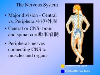
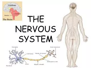
Nervous system: Central nervous system (CNS): brain and spinal cord Peripheral nervous system (PNS): cranial nerves and spinal nerves Two types of cells: neurons and supporting (glial) cells
Types of neurons: Interneuron- located entirely within CNS, integrates functions in CNS Sensory (from sensory receptor to CNS) Motor (from CNS to effector organ) somatic- stimulates skeletal muscles autonomic- affects smooth and cardiac muscle, also glandular secretion Nerve- bundle of nerves Ganglion- bundle of nerve cell bodies outside of CNS Nucleus- within CNS Tract- connects regions of CNS
Parts of a neuron Cell body- contains the nucleus and other organelles Dendrites- transmit electrical impulses TO the cell body Axon- transmits impulse AWAY from the cell body axons can be several feet long “Axon hillock” is located near the cell body nerve impulses originate there
Myelin is wrapped around the axon of many neurons In periphery, myelin is produced by Schwann cells In CNS, it is produced by oligodendrocytes
Structures of neurons sensory retina motor
Supporting cells Schwann cells, oligodendrocytes- produce myelin Satellite cells- support neurons in PNS Microglia-phagocytes in CNS Astrocytes- induces blood-brain barrier Ependymal cells- special epithelium that line brain ventricles and central canal of spinal cord also part of structure that makes CSF
Large axons are myelinated by Schwann cells or oligodendrocytes Gaps are left between the “wrappings” of each cell (nodes of Ranvier) Myelinated axons conduct nervous impulses more rapidly than unmyelinated In CNS, myelinated axons form “white matter” (Cell bodies and dendrites are gray matter)
Schwann cells can help repair damaged nerves Capacity for repair is much better in the periphery In fetal brain, neurotropins promote neuron growth Some factors help maintain neural structures in adult nervous systems Some inhibitory factors also
Astrocytes Most common glial cell in CNS Form blood-brain barrier Help with ion uptake Help with neurotransmitter uptake Many glucose transport carriers, which help move glucose from blood to brain
Blood-brain barrier (BBB) Probably due to effects of astrocytes on brain capillaries Everything must move into brain by diffusion and active transport Many substances (including therapeutic drugs) cannot cross BBB
Electrical activity in axons Resting membrane potential in neurons is –70 mV Large negatively charged molecules inside the cell Positively charged ions outside the cell (more Na out than K in) Neurons are excitable: they can change their membrane potential in response to stimulation
Permeability to ion changes Occurs in a very small area on the membrane Depolarization- potential difference approaches zero Repolarization- back to the resting potential Hyperpolarization- potential difference increases positive charges leave cell negative charges enter cell
Gated ion channels for K and Na (lots of these at axon hillock) Resting cell is more permeable to K than Na Depolarization- membrane becomes permeable to Na, and Na can diffuse into cell After Na gates close, K gates open and K diffuses out of the cell
Action potentials When completed, Na/K pumps restore balance of the ions Takes place on a very small part of membrane occurrence is rapid
Action potentials are very rapid Inactivation occurs until membranes are repolarized Stronger stimuli stimulate more and more axons (more action potentials are stimulated, but their amplitude does not change)
Refractory period When an action potential is being produced, a second stimulus will not affect that part of the membrane
Stimulus when K gates are open and membranes are repolarizing Relative refractory period- a very strong stimulus can overcome repolarizing
Conduction of nerve impulses Unmyelinated axon- wave of action potentials spreads along length of axon Amplitude of action potential does not change Myelinated axon- conduction rate is much faster
Graded potential Action potentials do not decrease in amplitude as they are conducted; graded are Graded potentials therefore signal only over a short distance postsynaptic potentials end-plate pacemaker, slow-wave
Graded vs action potentials All or none Magnitude varies with triggering event Propagated throughout membrane Decrement (can’t perpetuate itself) Triggered by depolari- zation to threshold Triggered by stimulus No refractory period Refractory period Occurs in specialized Occurs where there are Regions lots of Na+-gated channels
Synapse- connection between a neuron and a second cell From presynaptic to postsynaptic neuron Release of neurotransmitters (chemicals) A few electrical synapses in nervous system, In smooth muscle and heart gap junctions
Chemical synapses One-way Presynaptic neuron has synaptic vesicles Fusion of vesicles is mediated by calcium Calmodulin is activated Protein kinase activated Synaptic vesicles fuse with membrane Neurotransmitters diffuse across cleft and bind to receptors
Voltage-regulated channels in presynaptic axon Chemically regulated channels in postsynaptic membrane Ion channels are opened, depolarization occurs Can be excitatory or inhibitory Depends on which receptors are engaged Integration of impulses in dendrites and cell body of postsynaptic neuron
Acetylcholine Excitatory to some neurons in CNS and motor neurons Inhibitory to others Different cells have different types of receptors Nicotinic- stimulatory; nicotine also binds skeletal muscle fibers, autonomic ganglia Muscarinic- muscarine also binds smooth and cardiac muscle; glands
Ion channel most direct type of activation EPSP (excitatory postsynaptic potential) no threshold can be graded (number of stimulated receptors) no refractory period summation: effect of several EPSPs added (i.e., graded)
Muscarinic receptors- operated by G proteins Three subunits to G protein, different ones can be effectors Tends to have in inhibitory effect (IPSP)
Why inhibitory? K+ diffuses out, causing hyperpolarization Both EPSPs and IPSPs can be produced voluntarily- summate or cancel each other out
Acetylcholinesterase (AChE) Inactivates ACh. Otherwise ACh-receptor complexes would keep forming.
In PNS ACh stimulates muscles to contract In ANS: sympathetic and parasympathetic nerves Effect depends on whether nicotinic or muscarinic receptors are activated If EPSPs are above threshold an action potential will be generated along the axon
Monoamines as neurotransmitters Monoamines dopamine norepinephrine serotonin Tend to be stimulatory; must be quickly inhibited to maintain control
Control mechanisms: Uptake of monoamines by presynaptic neuron Degradation by monoamine oxidase in presynaptic neuron By post-synaptic neuron (COMT*, degrades catecholamines) COMT= catechol-O-methyltrasnferase
Monoamines act through second messenger (cAMP) Catecholamines norepinephrine- hormone and neuro- transmitter controlled by: reuptake monoamine oxidase (MAOIs inhibit this) COMT in postsynaptic neuron