Download
1 / 20
230 likes | 528 Vues
Respiratory Fungal Infections-II. Dr. Ahmed Al- Barrag Asst. Professor of Medical Mycology School of Medicine and the University Hospitals King Saud University. Candidiasis. Candidiasis refer to infection caused by any of the > 160 species of the genus Candida Etiology: Candida species

E N D
Respiratory Fungal Infections-II Dr. Ahmed Al-BarragAsst. Professor of Medical MycologySchool of Medicine and the University HospitalsKing Saud University
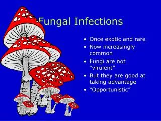
Candidiasis • Candidiasisrefer to infection caused by any of the > 160 species of the genus Candida • Etiology: Candida species • Yeasts • Pseudohyphae • Candida albicans is commonly responsible for candidiasis. • Other species include: • Candida glabrata, • Candida tropicalis, • Candida parapsilosis, • Candida krusei
Candida Part of the endogenous flora: Skin Gut Mucosal surfaces Most infections are due to person’s own flora Portal of entry: Breach in skin or mucosa by catheters, trauma, surgery Endogenous source for majority of Candida infections Exogenous transmission?
Candidiasis High-risk patients AIDS Surgery Malignancy Burns Premature infants • Exposures • ICU >7 days • CVCs • Antibiotics • TPN • Colonization
Candidiasis Disease spectrum • Infections of the skin and nail • Gastrointestinal infections (oral cavity, esophagus) • Infections of genitalia (female) • Urinary tract infection (lower and upper UTIs) • Ocular infections (Keratitis, endophthalmitis) • Candidemia • CNS infection • Deep organ Candidiasis • Pneumonia • Endocarditis • Bone and joint infections • Chronic mucocutaneouscandisiasis (CMC) (congenital, immunological defect)
Pulmonary Candidiasis Primary pneumonia is less common and could be a result of Aspiration Secondary pneumonia commonly seen with hematogenouscandisiasisImmunocompromised patients Diagnosis: Isolation of Candida from sputum, BAL is not always significant Radiology, clinical features Lung biopsy Other yeast causing Pulmonary infections Trichosporon Geotrichum
Candidemia Increased colonization (endogenous or exogenous factors) Damage in host barriers by catheters, trauma, surgery Immunosuppression Central venous catheters (CVC) Disseminated candidiasis (envolment of any organ) Septic shock Meningitis Ocular involvement (retinitis)
Candida- Nosocomial Bloodstream Infections Candida is the fourth in causing nosocomial bloodstream infections (BSI) WisplinghoffH, et al. Clin Infect Dis. 2004;39:309-317.
Candidiasis - diagnosis Laboratory Diagnosis: Specimen depend on the site of infection. Swabs, Urine, Blood, Respiratory specimens, CSF, Blood for serology Direct microscopy : Gram stain, KOH, Giemsa, GMS, or PAS stained smears. Budding yeast cells and pseudohyphae will be seen in stained smear or KOH.
Candidiasis - diagnosis Culture: on SDA & Blood agar at 37oC, creamy moist colonies in 24 - 48 hours. Blood culture
Candidiasis - diagnosis Because C. albicansis the most common species to cause candidiasis We do the following tests to identify C. albicans 1. Germ tube test Formation of germ tube when cultured in serum at 37ᵒC 2. Chlamydospore production in corn meal Agar (CMA) Germ tube test 3. Resistance to 500 μg/ml Cycloheximide(will grow on Mycobiotic Medium) If these 3 are positive yeast is C.albicans, • If negative, then it could be any other yeast, • Use Carbohydrate assimilations and fermentation. Chlamydospores of C. albicansin CMA • commercial kits available for this like: API 20C, API 32C • Culture on Chromogenic Media (CHROMagar™ Candida)
Candidiasis - diagnosis Serology: Patient serum Test for Antigen , e.g. Mannan antigen using ELISA Test for Antibodies PCR
Treatment of Candidiasis Systemic treatment of Candidiasis: Fluconazole Voriconazole Caspofungin Amphotericin • Candidemia: • Treat for 14 days after last positive culture and resolution of signs and symptoms • Remove all intravascular catheters, if possible
In Vitro Susceptibility of Candida spp. Antifungal susceptibility testing in not done routinely in the microbiology lab. • Points to consider: • C. glabratacan be less susceptible or resistant to fluconazole C. kruseiis resistant to fluconazole
Pulmonary Cryptococcosis • Causative agent • Cryptococcus neoformans • C. gattii • A typical yeast with a thick capsule • Source of infection • Pigeon or birds droppings & contaminated soil • Pathogenesis • Human infection by inhalation • infections could be asymptomatic • May develop pneumonia, disseminate to CNS causing meningitis (immunological status of the host)
Cryptococcosis Lab Diagnosis 1. India Ink preparation Yeast cell with a thick capsule 2. Culture on SDA Identify using API 20C , Urease +ve Phenol oxidase +ve 3. Serology : Capsular Antigen by latex agglutination excellent sensitivity
Cryptococcosis Treatment Systemic fungal agents Amphotericin B • Combination of Amphotericin B & flucytosine
Pneumocystosis(PCP) Opportunistic fungal pneumonia It is interstitial pneumonia of the alveolar area. Affect compromised host Especially common in AIDS patients. Etiology: Pneumocystisjiroveci • Previously thought to be a protozoan parasite. • It has been proven to be a fungus • Does not grow in laboratory media e.g. SDA • Naturally found in rodents (rats), other animals (goats, horses), Humans contract it during childhood.
Pneumocystosis • Laboratory Diagnosis: • Patient specimen: Bronchoscopic specimens (B.A.L.), Sputum, Lung biopsy tissue. • Histology sections or smears stained by Silver stain (GMS). better sensitivity)) Immunuofluorescence If positive will see cysts of hat-shape, cup shape, crescent Treatment: Trimethoprim – sulfamethoxazole